
Table 1
Hemodynamic parameters of cardiac catheterization and cardiac magnetic resonance imaging. PA: pulmonary artery; PCWP: pulmonary capillary wedge pressure; PVR: pulmonary valve regurgitation; RA: right atrium; RV: right ventricle; RVEDVi: right ventricular end-diastolic volume index; TVR: tricuspid valve regurgitation.
| CARDIAC CATHETERIZATION | CARDIAC MAGNETIC RESONANCE IMAGING | ||||||||
|---|---|---|---|---|---|---|---|---|---|
| RA (mm Hg) | RV (mm Hg) | PA (mm Hg) | PVR (Wu) | PCWP (mm Hg) | Shunt Fraction (Qp/Qs) | RVEDVi (mL) | TVRSeverity | PVR Severity | |
| Case 1 | 19 | 44/23 | 30/20 | 1.3 | 18 | 0.9 | 152 | Mild | Moderate to severe |
| Case 2 | 12 | 45/10 | 45/12 | 2 | 16 | 0.93 | 165 | Moderate | Severe |
| Case 3 | 20 | 72/20 | 60/30 | 2.5 | 22 | 0.77 | 207 | Severe | Severe |
| Case 4 | 13 | 27/9 | 28/10 | 1 | 14 | 0.69 | 143 | Moderate | Severe |
Video 1
Main pulmonary angiography demonstrating degenerated right ventricle-pulmonary artery homograft with pulmonic regurgitation; also at https://youtube.com/shorts/10_v97YnH3Q.
Video 2
Main pulmonary angiography demonstrating prosthetic pulmonic valve competence following implantation of Palmaz XL P4010 stent and Melody valve; also at https://youtube.com/shorts/Fnbif5eio0s.
Video 3
Intracardiac echocardiography performed from the right atrium with color Doppler demonstrating right to left shunt through the PFO at baseline; also at https://youtu.be/OOFEhr55Sgg.
Video 4
Intracardiac echocardiography demonstrating no residual right to left shunt through the PFO following implantation of a 25mm Amplatzer PFO Occluder (Abbott, Santa Clara); also at https://youtu.be/xBEzK-gxYK4.
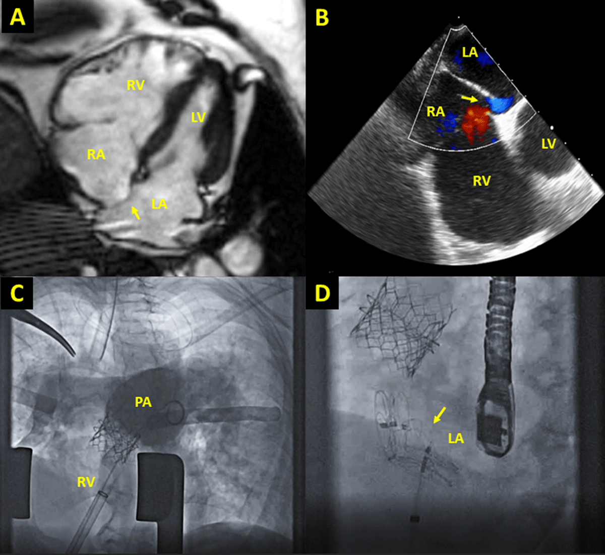
Figure 1
Case imaging. (A) Four-chamber cardiac magnetic resonance imaging demonstrating severely dilated RV compared to LV with leftward bowing of interatrial septum (arrow). (B) Transesophageal echocardiography demonstrating right-to-left shunt through PFO (arrow). (C) Fluoroscopic images from hybrid pulmonary valve replacement and PFO closure; angiogram demonstrates no residual pulmonic regurgitation. (D) Two occluders (arrow) were deployed to close the PFO and atrial septal deflect. LA: left atrium; LV: left ventricle; PA: pulmonary artery; PFO: patent foramen ovale; RA: right atrium; RV: right ventricle.
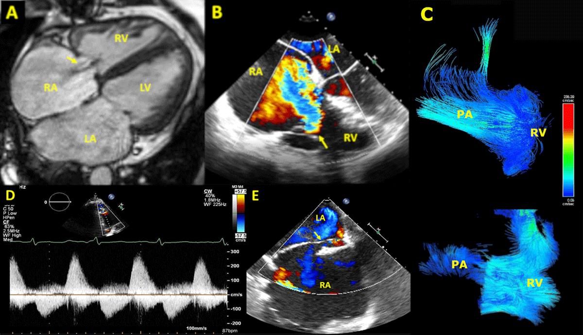
Figure 2
Severe PV regurgitation leading to severe functional TV regurgitation, favoring a right-to-left shunt via PFO. Severe TV regurgitation jet shown by 4-chamber view (A) CMR and (B) transesophageal echocardiography. (C) Four-dimensional CMR demonstrating severe PV regurgitation. (D) Spectral doppler showing classic PV regurgitation waveform. (E) PFO with right-to-left shunt by transesophageal echocardiography. LA: left atrium; LV: left ventricle; PA: pulmonary artery; PFO: patent foramen ovale; PV: pulmonary valve; RA: right atrium; RV: right ventricle; TV: tricuspid valve.