
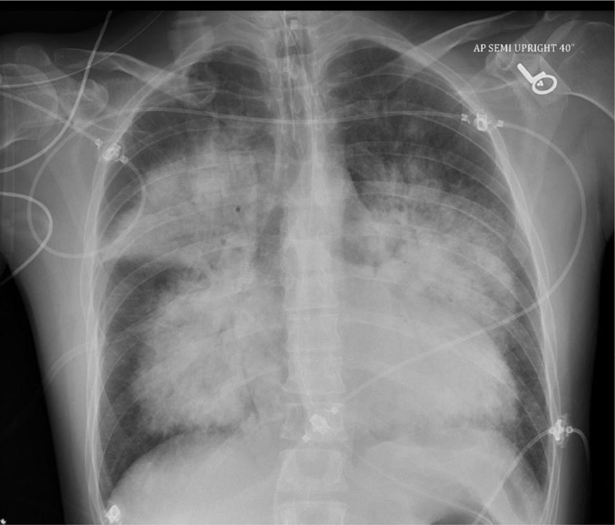
Figure 1
Chest x-ray upon presentation showing extensive dense perihilar mass-like consolidative opacities.
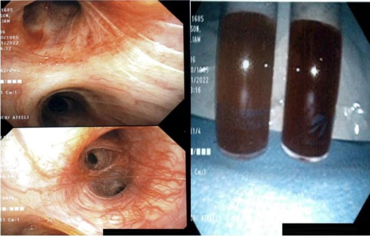
Figure 2
Bronchoscopy showing alveolar hemorrhage.
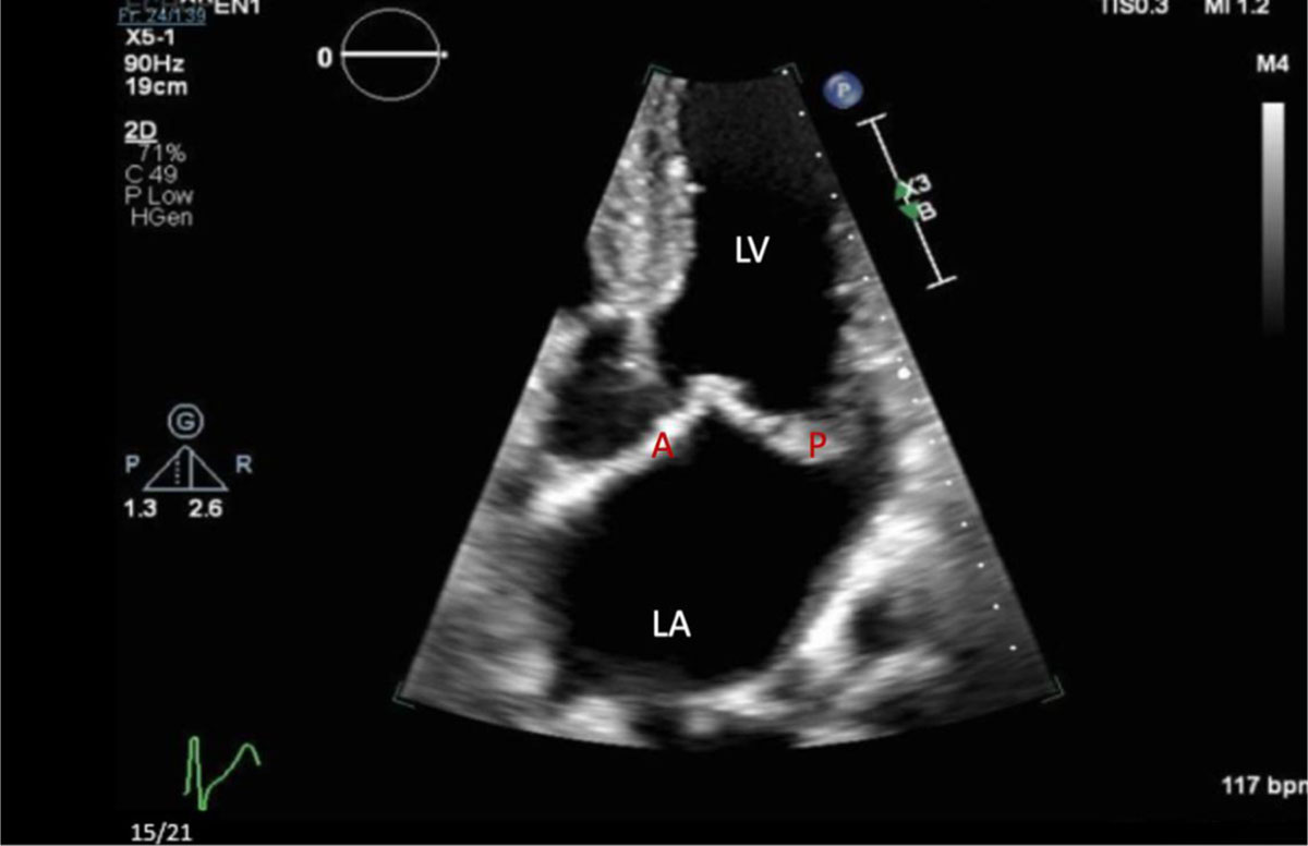
Figure 3
Zoomed in transthoracic echocardiogram with apical four-chamber view showing the left ventricle (LV), left atrium (LA), posterior (P), and anterior (A) mitral valve leaflets.
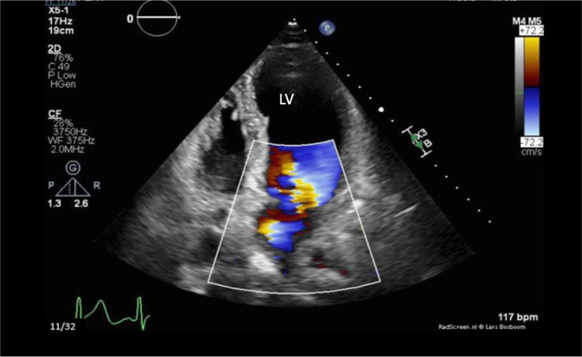
Figure 4
Transthoracic echocardiogram with apical four-chamber view in systole, showing severe jet of mitral valve regurgitations.
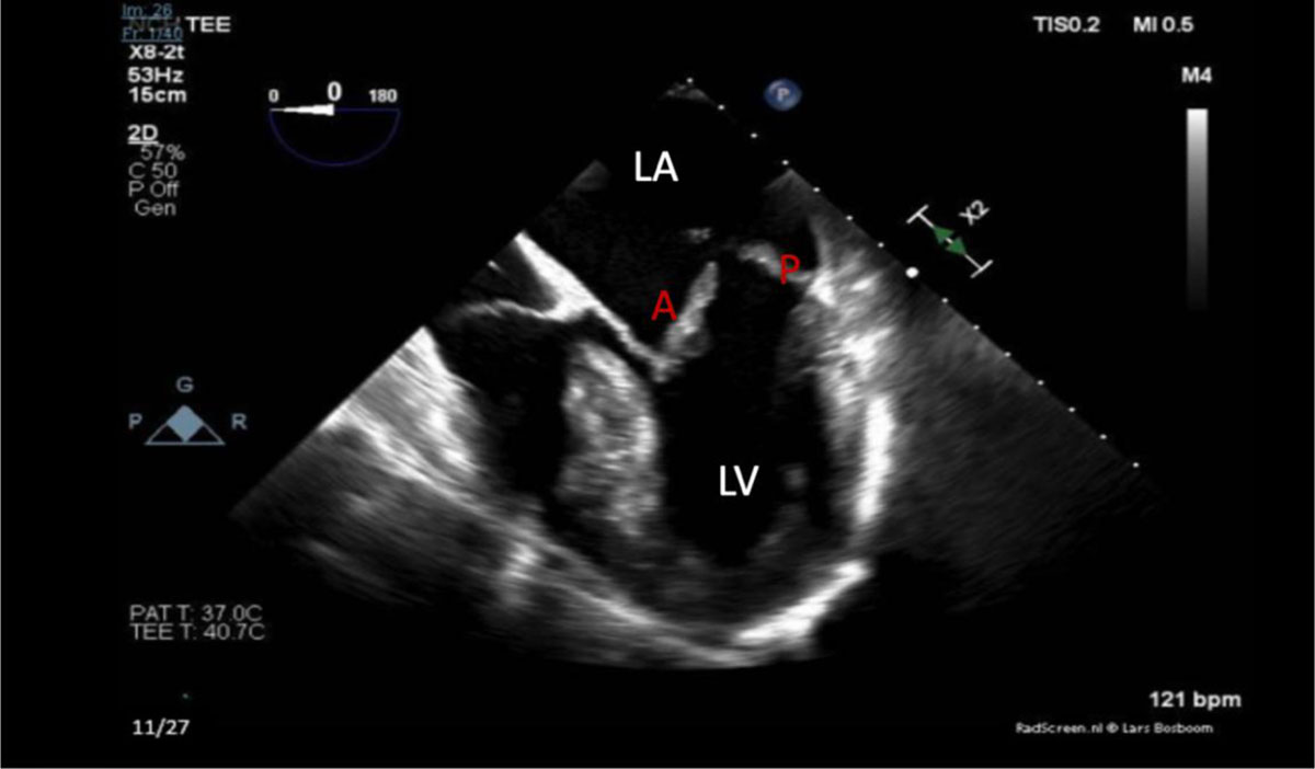
Figure 5
Mid-esophageal transesophageal echocardiogram with 4-chamber apical view view showing left ventricle (LV) and left atrium (LA). Myxomatous degeneration and severe prolapse of the anterior and posterior leaflets of the mitral valve are seen on this image.
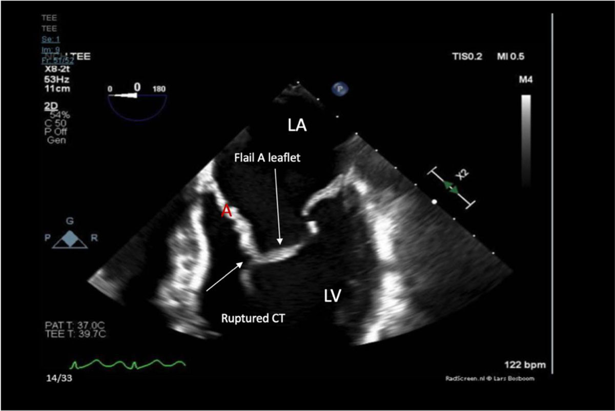
Figure 6
Zoomed in mid esophageal transesophageal echocardiogram with 4-apical view showing left ventricle (LV), left atrium (LA), chordae tendineae (CT), and flail (A) mitral valve leaflet.
Video 1
Transesophageal echocardiograms showing a myxomatous mitral valve with anterior leaflet prolapse along with chordal rupture involving the flail anterior leaflet, see also at https://youtu.be/J6KtVU3eSNk.
Video 2
Color Doppler transesophageal echocardiogram showing the mitral valve with flail anterior leaflet and severe mitral regurgitation, see also at https://youtu.be/pSyQjq4Xm4A.
Video 3
Zoomed 3-dimensional transesophageal echocardiogram view of the myxomatous mitral valve showing the flail anterior leaflet, see also at https://youtu.be/d1v8SeTqmmE.