
Figure 1
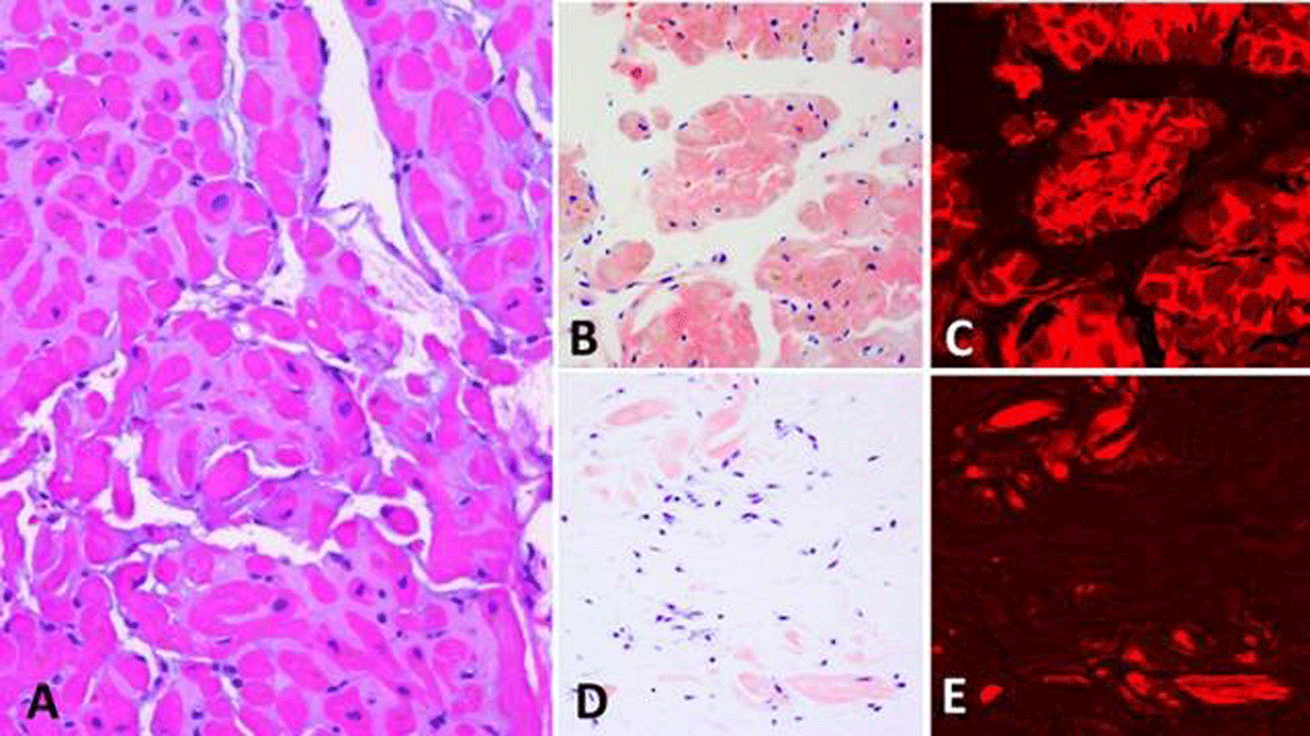
(A-C) Cardiac amyloidosis. (A) Amyloid appearing as a glassy eosinophilic material surrounding myocardial fibers (Hematoxylin&eosin ×200). (B, C) Amyloid displaying a “salmon-pink” color under Congo red stain (×200), which is strongly enhanced under Texas red immunofluorescent examination (×200). (D, E) Tenosynovial amyloidosis. A small amount of amyloid not apparent in routine tissue section appearing here as small “salmon-pink” nodules, which are strongly enhanced under Texas red immunofluorescent examination (×200). Even this small amount of amyloid is suitable for mass spectrometric analysis; in this case it shows transthyretin type of amyloid. Transthyretin amyloid identified in tissue removed for carpal tunnel syndrome may signify concurrent or future cardiac amyloidosis of the same chemical type.
Table 1
Revised Mayo Clinic staging of AL amyloidosis (with chemotherapy). Scores are calculated by giving 1 point for each of the following: troponin T ≥ 0.025 ng/ml; N-T pro BMP ≥ 1,800 pg/ml; difference between involved and uninvolved serum free light chain levels >180 mg/L.
| SCORE | MEDIAN SURVIVAL (MONTHS) |
|---|---|
| 0 | 94 |
| 1 | 40 |
| 2 | 14 |
| 3 | 6 |
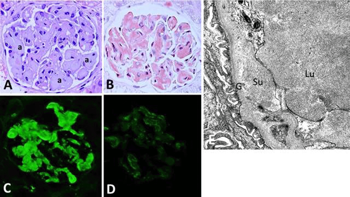
Figure 2
Renal amyloidosis. (A) Amyloid appearing as glassy material (a) deposited along glomerular capillary wall and lumens with hematoxylin &eosin stain, which (B) appears as “salmon-pink” deposits with Congo red stain. (C) The amyloid is composed of kappa light chain (D) but negative for lambda light chain, indicating light chain type of amyloid (×400 for panels A–D). (E) By electron microscopy (×15,000), amyloid appearing as short non-branching fibrils in the subendothelial location and glomerular capillary lumen. Su: subendothelial; Lu: glomerular capillary lumen; G: glomerular basement membrane; Po: podocyte