
Table 1
Definitions of contrast-associated acute kidney injury. KDIGO: Kidney Disease Improving Global Outcomes; NCDR: National Cardiovascular Data Registry-Acute Kidney Injury; AKIN: Acute Kidney Injury Network; RIFLE: Risk, injury, failure, loss of kidney function, and end-stage kidney disease; ESUR: European Society of Urogenital Radiology.
| KDIGO (also used by NCDR) | Increase in serum creatinine of ≥ 0.3 mg/dL within 48 hours or ≥ 50% within 7 days, or urine output of < 0.5 mL/kg/hour for > 6 hours |
| AKIN | Increase in serum creatinine of ≥ 0.3 mg/dL or ≥ 50% within 48 hours, or urine output of < 0.5 mL/kg/hour for > 6 hours |
| RIFLE | Risk: Increase in serum creatinine to 1.5 times baseline, or urine output of < 0.5 mL/kg/hour for 6 to 12 hours Injury: Increase in serum creatinine of up to 2 times baseline, or urine output of < 0.5 mL/kg/hour for 12 to 24 hours Failure: Increase in serum creatinine to 3 times baseline, or increase in serum creatinine by > 0.5 mg/dL to > 4.0 mg/dL, or urine output of < 0.3 mL/kg/hour for > 24 hours or anuria for > 12 hours, or initiation of kidney replacement therapy Loss: Need for kidney replacement therapy for > 4 weeks End stage: Need for kidney replacement therapy for > 3 months |
| ESUR | Increase in serum creatinine by more than 25% or 0.5 mg/dL within 3 days following the intravascular administration of a contrast medium in the absence of an alternative etiology |
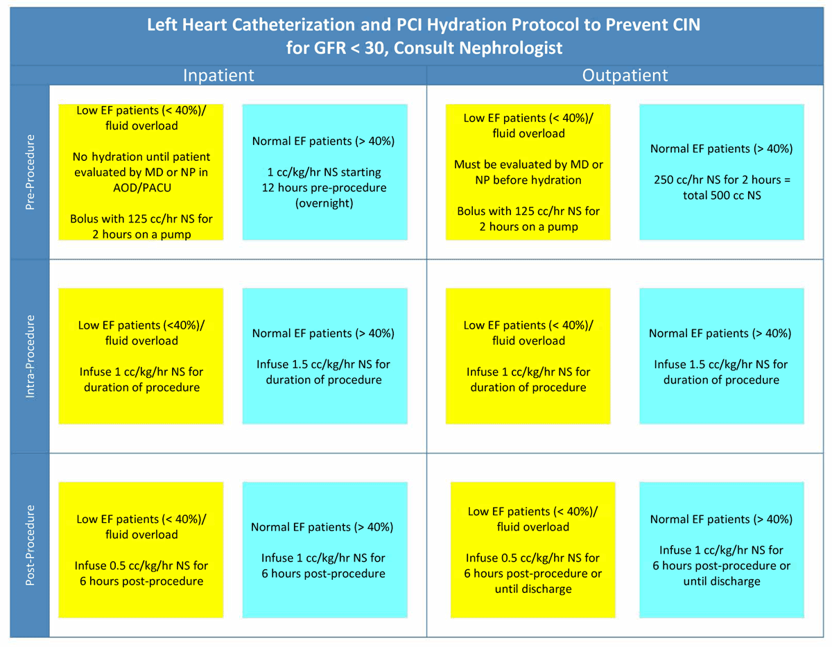
Figure 1
Houston Methodist Cardiac Catheterization Lab hydration protocol to prevent contrast associated acute kidney injury. EF: ejection fraction; PACU: post-anesthesia care unit; NS: normal saline
Table 2
Contrast-sparing strategies recommended for all percutaneous coronary interventions when possible but especially in the presence of chronic kidney disease.
| Angiography | Retrieve previous (and recent) diagnostic coronary angiograms to avoid repeat acquisition. |
| Use high frame rate (eg, 30 frames/s) acquisitions to improve image quality (at the cost of higher radiation dose). | |
| Consider biplane angiography. | |
| Guiding catheters | Avoid side-holes in guide catheters where possible. |
| Avoid test injections with contrast to determine guide catheter engagement. Instead, use a coronary wire or inject normal saline (EKG repolarization changes confirm guide engagement). | |
| Contrast media and volume | Use iso-osmolal or low-osmolal contrast media (In practice, high-osmolal agents are rarely if ever used for coronary angiography). |
| Limit or eliminate the volume of contrast per injection (2 mL/injection). | |
| Use automated contrast injectors. | |
| Use diluted (with 50% normal saline) contrast media. | |
| Eliminate contrast in the guide catheter by back bleeding prior to administration of medications or advancing equipment. | |
| In high-risk patients, consider the use of newer devices that minimize contrast injection volumes or divert contrast from the kidney (see details below). | |
| Vessel wiring, lesion assessment, stent deployment and optimization | Use a previously performed coronary computed tomography angiogram to create a live road map of the coronary tree for guidewire navigation (Syngo Fusion, Seimens Healthcare; SmartCT Roadmap, Phillips). |
| Wire side branches (metallic roadmap) to aid optimal stent positioning. | |
| Use instant wave-free ratio to evaluate the hemodynamic significant of the lesion(s) (ie, is intervention truly needed?). | |
| Use intravascular ultrasound or dextran-based optical coherence tomography (experimental) to locate and assess lesions, identify proximal and distal stent landing zones, and confirm adequate stent expansion and apposition. | |
| Use stent enhancement technologies to position balloons and confirm adequate expansion (ClearStent, Siemens Healthcare; StentBoost, Philips; Intrasight Device Detection, Philips). |