
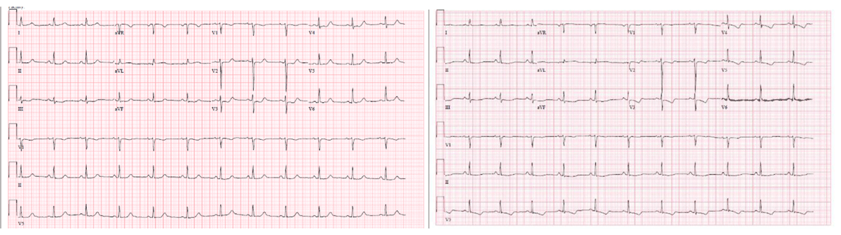
Figure 1
Echocardiography showing dynamic ST/T wave changes.

Figure 2
(A, B) Spontaneous coronary artery dissection of diagonal branch of the bifid left anterior descending coronary artery. (C) Normal caliber ramus coronary artery.
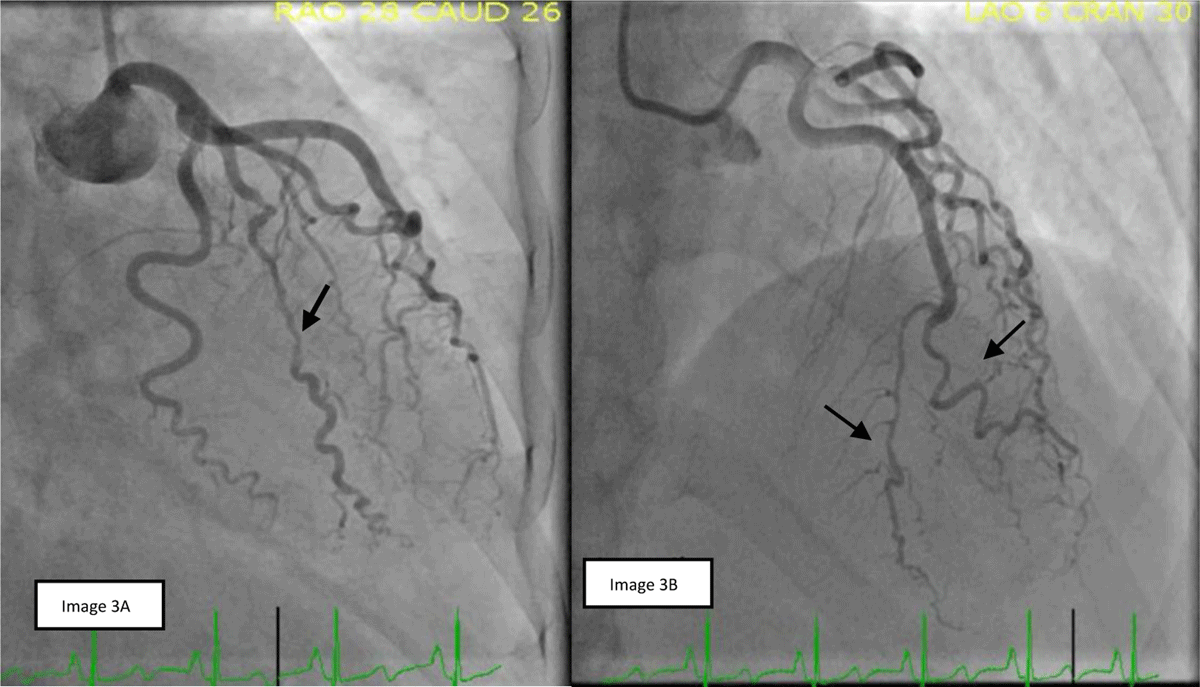
Figure 3
(A) Spontaneous coronary artery dissection (SCAD) of ramus coronary artery. (B) Possible SCAD in the apical branch of bifid left anterior descending coronary artery (LAD) and healing of previous SCAD in diagonal branch of bifid LAD.
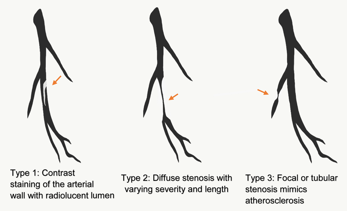
Figure 4
Diagnosis of spontaneous coronary artery dissection is based on coronary angiography, with appearance classified into three Yip-Saw types.