
Video 1
Marantic endocarditis echocardiogram, also at https://youtu.be/akg4UiTxh4M.
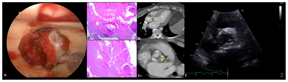
Figure 1
Marantic endocarditis. (A) Intraoperatively, the valve appears stenotic with a heavily calcified annulus and fresh-formed thrombus on both the outflow and inflow. (B) Low magnification of a vegetation composed of fibrin, forming an eosinophilic mass. Mature fibrosis is seen at the base of the vegetation. (C) High magnification shows that vegetation is composed predominantly of fibrin and red blood cells. (D) 3-chamber view on non-gated computed tomography shows ventricular hypertrophy and subvalvular aortic valve thickening extending to the left ventricular outflow tract (arrows). (E) Short axis of the aortic valve on non-gated computed tomography shows bicuspid aortic valve opening (Sievers type 0) with severe thickening of the left coronary cusp leaflet and no calcification (arrows). (F) Parasternal short axis view shows thickened aortic valve leaflets with systolic doming and restricted opening.