
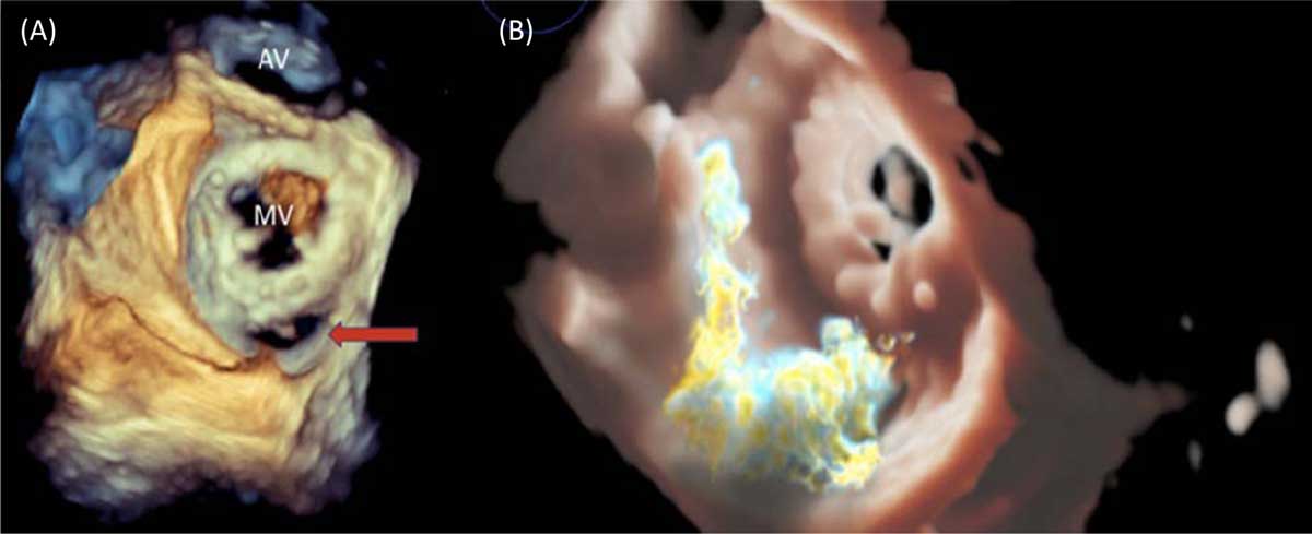
Figure 1
(A) 3-dimensional (3D) enface view of the mitral valve (MV) with the aortic valve (AV) seen in the 12 o’clock position. The mitral bioprosthesis is visualized with a paravalvular defect located posteromedially (red arrow). (B) 3D Trueview image of the mitral valve showing eccentric systolic flow through the paravalvular defect.
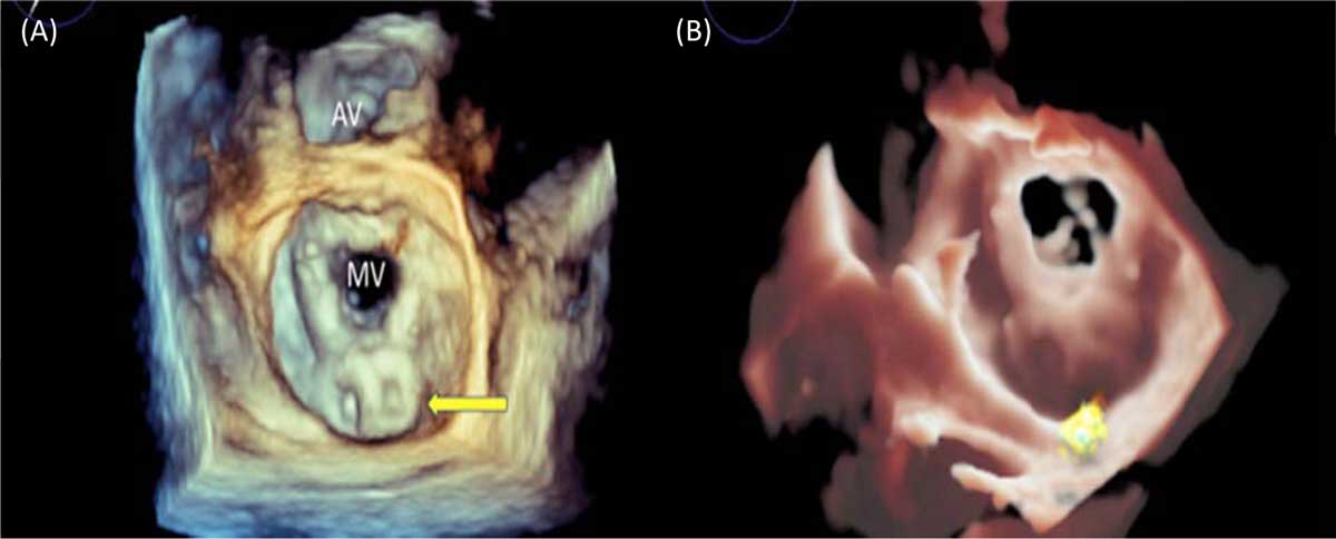
Figure 2
(A, B) A 3-dimensional enface view of the mitral valve with two Amplatzer ventricular septal defect (Abbott) devices (yellow arrow) well seated within the paravalvular defect. Panel B shows a small amount of residual paravalvular leak. AV: aortic valve; MV: mitral valve
Competing Interests
The authors have no competing interests to declare.