
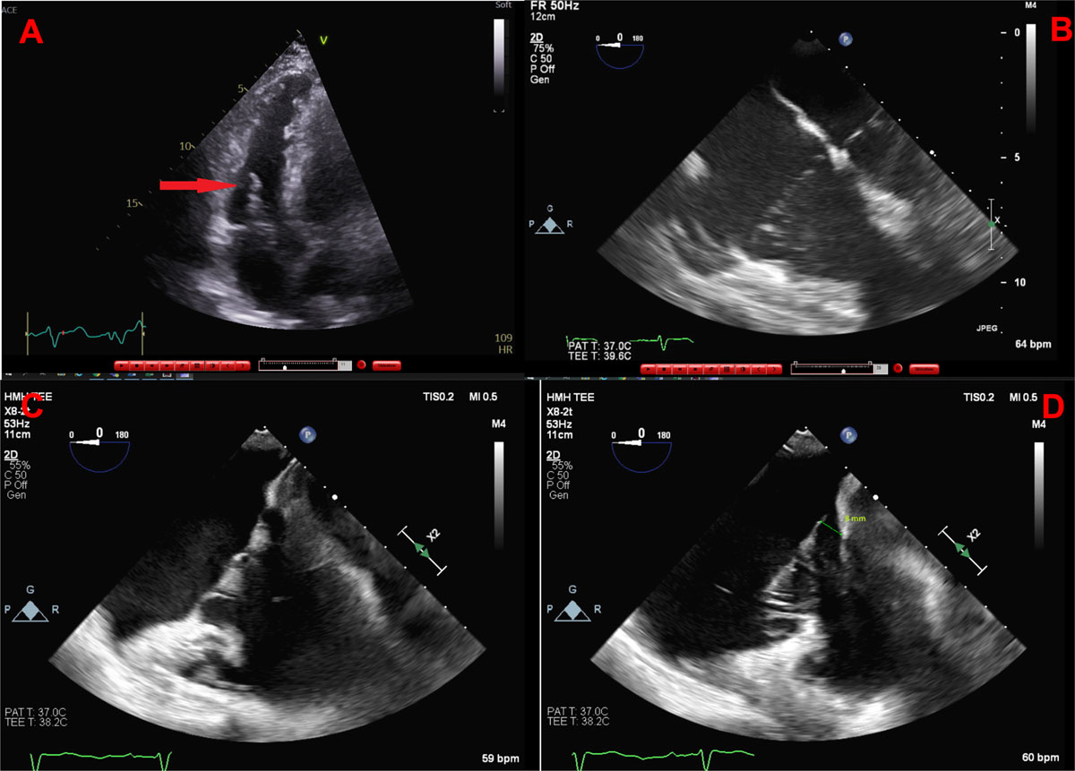
Figure 1
(A) Transthoracic echocardiography image demonstrates an apparent echo density on the tricuspid valve. (B) Transesophageal echocardiography (TEE) image demonstrates tricuspid prolapse with the mitral and tricuspid valves at equal level in systole. (C) TEE image shows the tricuspid leaflets in early systole at the level of the annulus. (D) TEE image shows the tricuspid leaflets in late systole prolapsing 8 mm into the right atrium.