
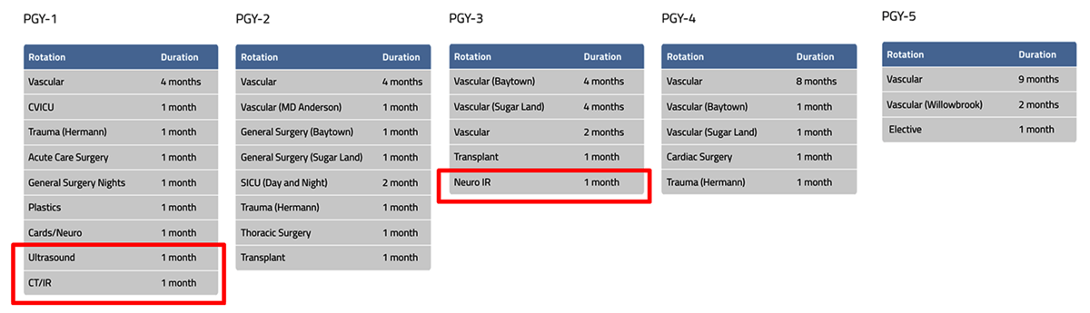
Figure 1
Integrated 0+5 vascular surgery residency curriculum; red boxes indicate training rotations related to imaging.
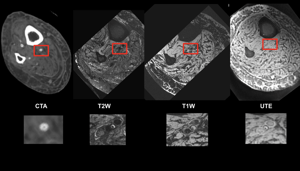
Figure 2
Tibial vessels imaged with computed tomography angiography (CTA) and magnetic resonance imaging (MRI) using T2-weighted, T1-weighted, and ultrashort echo time sequences. The CTA shows concentric calcium but suffers from beam hardening artifacts and calcium blooming that obscures the vessel lumen. MRI with ultrashort echo time imaging shows a very thin rim of hypointensity around the central vessel, indicating that the calcium ring is thin and the lumen is occluded with mixed morphology plaque.11
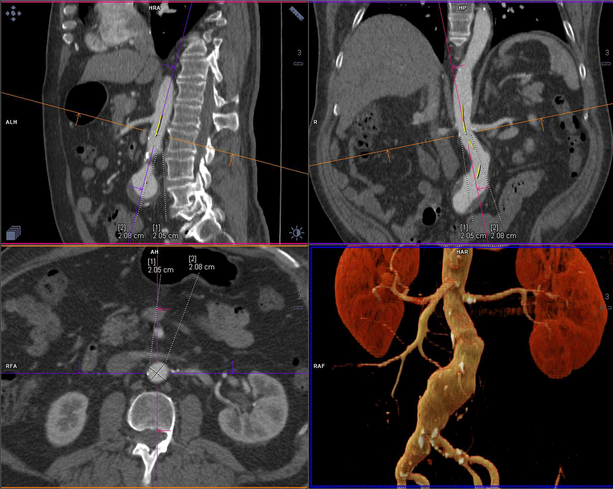
Figure 3
Three-dimensional multiplanar reconstruction determines aortic neck diameter, angle, and centerline measurements to size abdominal aortic endograft.
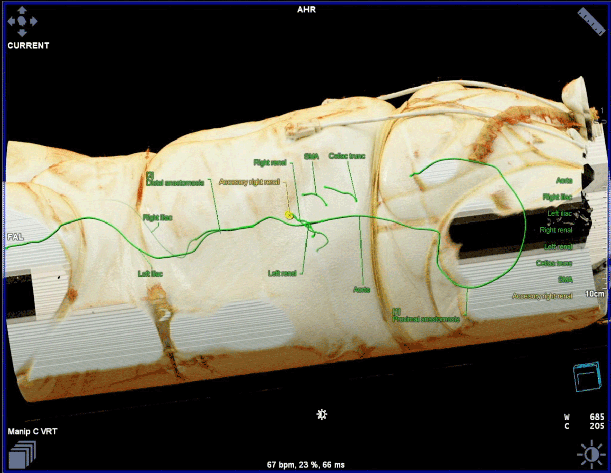
Figure 4
Cinematic rendering helps plan the optimal incision and approach for open repair of type 4 thoracoabdominal aneurysm.11, 13
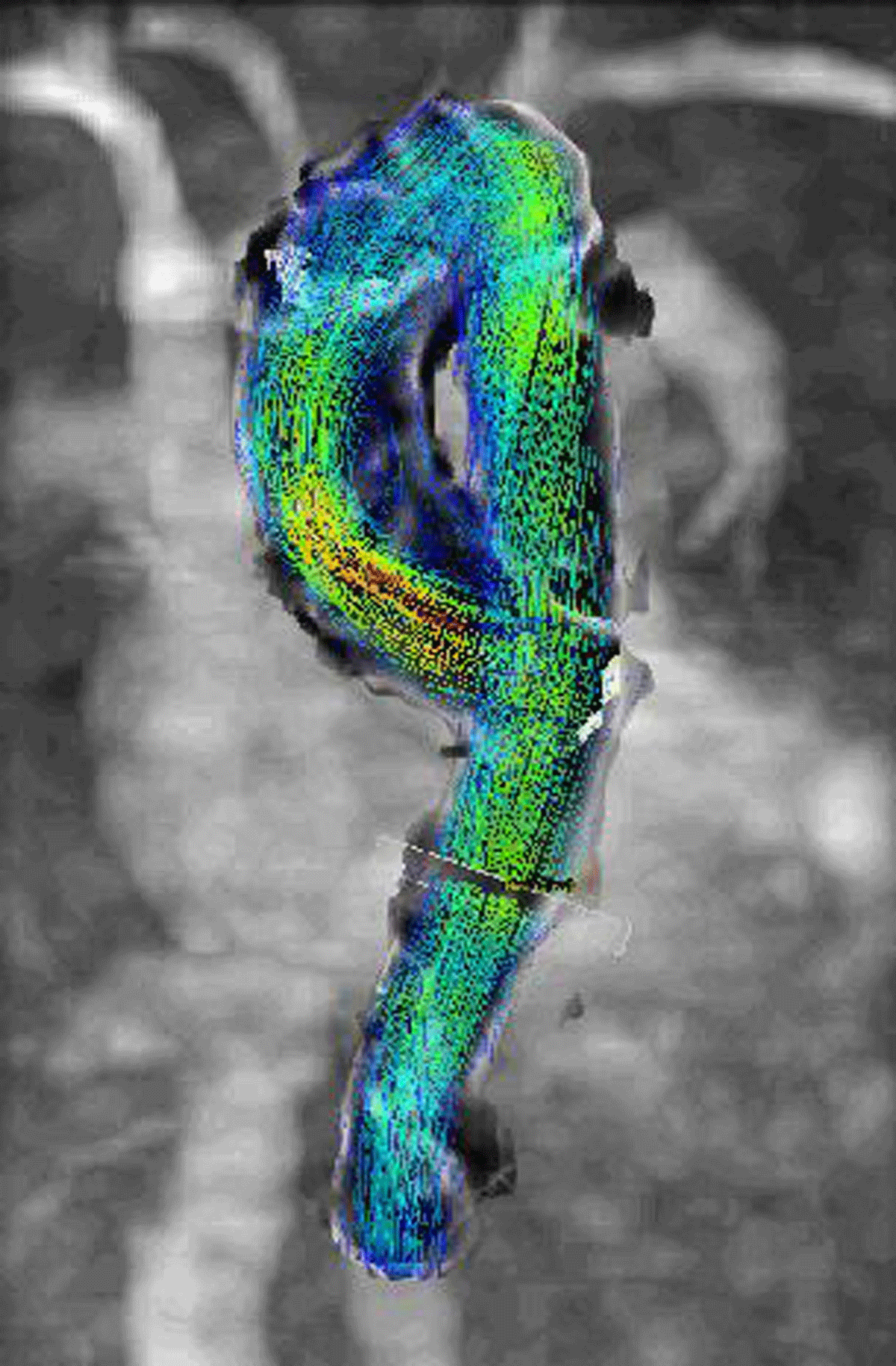
Figure 5
An example of four-dimensional (4D) flow magnetic resonance imaging, which enables the visualization of the temporal evolution of complex blood flow patterns within an acquired 3D volume. The blood flow patterns are represented in the colored vectors, which represent velocity with blue being closer to 0 m/s and red up to 1 m/s.17, 18, 19
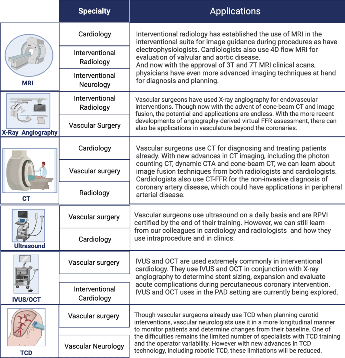
Figure 6
The strengths and capabilities of an interdisciplinary team in an advanced vascular imaging fellowship demonstrate the various applications of the different imaging modalities.22, 23 MRI: magnetic resonance imaging; CT: computed tomography; IVUS/OCT: intravascular ultrasound/optical coherence tomography; FFR: fractional flow reserve; CTA: CT angiography; RPVI: Registered Physician in Vascular Interpretation; PAD: peripheral artery disease; TCD: transcranial doppler
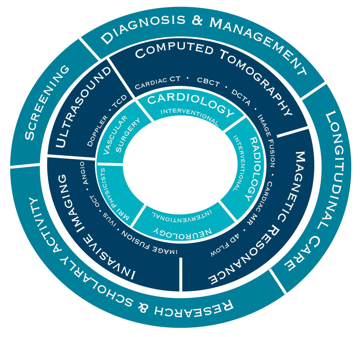
Figure 7
The core competencies of our proposed advanced vascular imaging fellowship. TCD: transcranial doppler; CBCT: cone-beam computed tomography; DCTA: dynamic CT angiography; IVUS: intravascular ultrasound; OCT: optical coherence tomography