
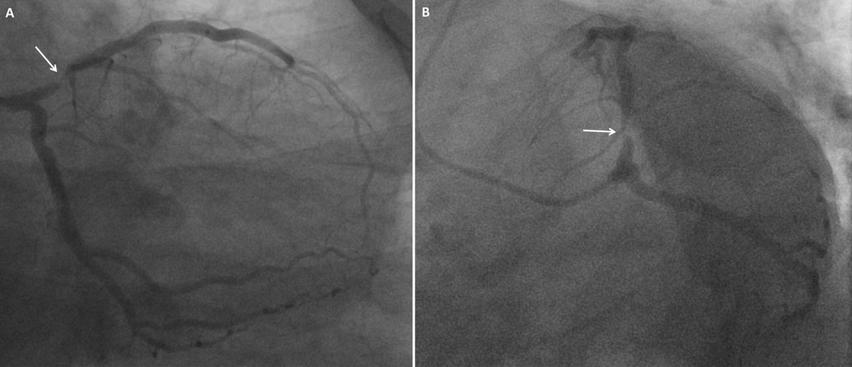
Figure 1
(A) RAO caudal view and (B) LAO caudal view showing a tubular 90% stenosis in the proximal LAD artery (arrow) and unremarkable LCx artery. RAO: right anterior oblique; LAO: left anterior oblique; LAD: left anterior descending; LCx: left circumflex.
Video 1
LAO caudal view, baseline angiogram of the left coronary arteries showing a tubular 90% stenosis in the proximal LAD artery and unremarkable LCx artery; also at https://youtube.com/shorts/BQNZMUK7jZ4. LAO: left anterior oblique; LAD: left anterior descending; LCx: left circumflex.
Video 2
LAO cranial view, baseline angiogram of the left coronary arteries showing no significant coronary artery disease in the mid to distal segments of the LAD and LCx coronary arteries; also at https://youtube.com/shorts/Cdq5XpylD_Y. LAO: left anterior oblique; LAD: left anterior descending; LCx: left circumflex.
Video 3
LAO view, baseline angiogram of the right coronary artery with no significant coronary artery disease; also at https://youtu.be/kqfThoyTGuI. LAO: left anterior oblique.
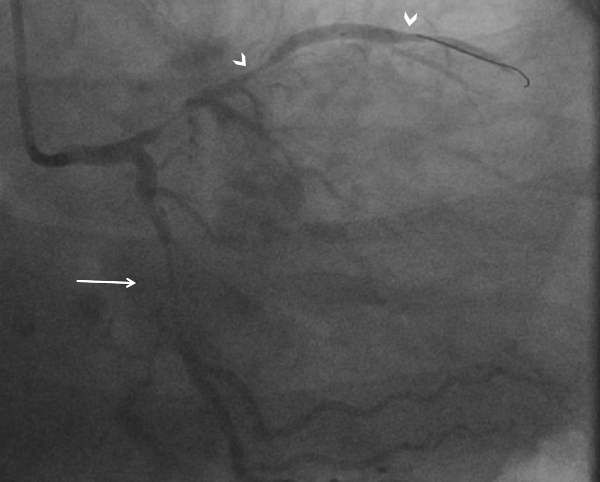
Figure 2
RAO caudal view showing diffuse stenosis in the proximal to mid segment of the previously unremarkable LCx artery with TIMI 2 flow (arrow) and diffuse stenosis in the previously normal mid to distal LAD segments with TIMI 0 flow (arrowheads), consistent with vasospasm. RAO: right anterior oblique; LCx: left circumflex; LAD: left anterior descending; TIMI: thrombolysis in myocardial infarction.
Video 4
RAO caudal view showing diffuse stenosis in the proximal to mid segment of the previously unremarkable LCx artery with TIMI 2 flow and diffuse stenosis in the previously normal mid to distal LAD segments with TIMI 0 flow, consistent with vasospasm; also at https://youtube.com/shorts/Da8UVUjPBik. RAO: right anterior oblique; LCx: left circumflex; TIMI: thrombolysis in myocardial infarction; LAD: left anterior descending.

Figure 3
RAO caudal view showing diffuse stenoses in the LAD and LCx arteries with angiographically different segment involvement and severity. Compared to Figure 2, there is shorter segment involvement of the mid LCx artery (arrow) and more severe involvement of the mid to distal LAD artery (arrowheads). RAO: right anterior oblique; LAD: left anterior descending; LCx: left circumflex.
Video 5
RAO caudal view showing diffuse stenoses in the LAD and LCx arteries with angiographically different segment involvement and severity. Compared to Figure 2 or Video 4, this shows a shorter segment involvement of the mid LCx artery and more severe involvement of the mid to distal LAD artery; also at https://youtube.com/shorts/80DlJSbQJB4. RAO: right anterior oblique; LAD: left anterior descending; LCx: left circumflex.
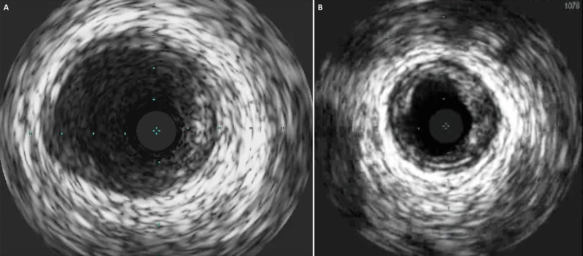
Figure 4
IVUS of the (A) left main coronary artery showing absence of dissection and (B) LCx artery showing absence of dissection with mild to moderate atherosclerotic disease. IVUS: intravascular ultrasound; LCx: left circumflex.
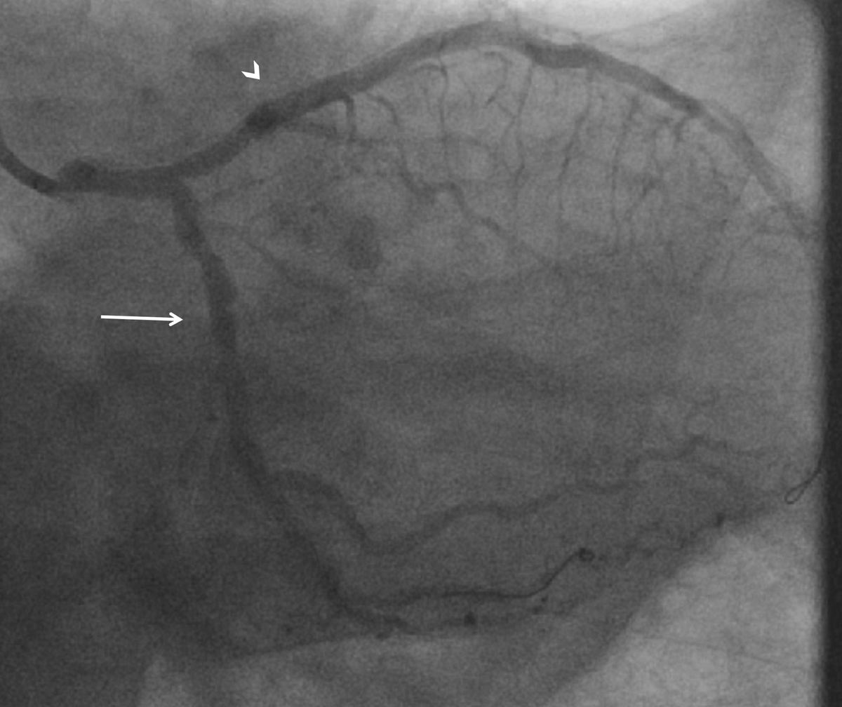
Figure 5
RAO caudal view showing resolution of LCx artery spasm (arrow) and presence of newly placed stents in the LAD artery (arrowhead) with TIMI 3 flow in both arteries. RAO: right anterior oblique; LCx: left circumflex; LAD: left anterior descending; TIMI: thrombolysis in myocardial infarction.
Video 6
Final result. RAO caudal view showing resolution of LCx artery spasm and presence of newly placed stents in the LAD artery with TIMI 3 flow in both arteries; also at https://youtube.com/shorts/BKSP7L-wXG0. RAO: right anterior oblique; LCx: left circumflex; LAD: left anterior descending; TIMI: thrombolysis in myocardial infarction.