
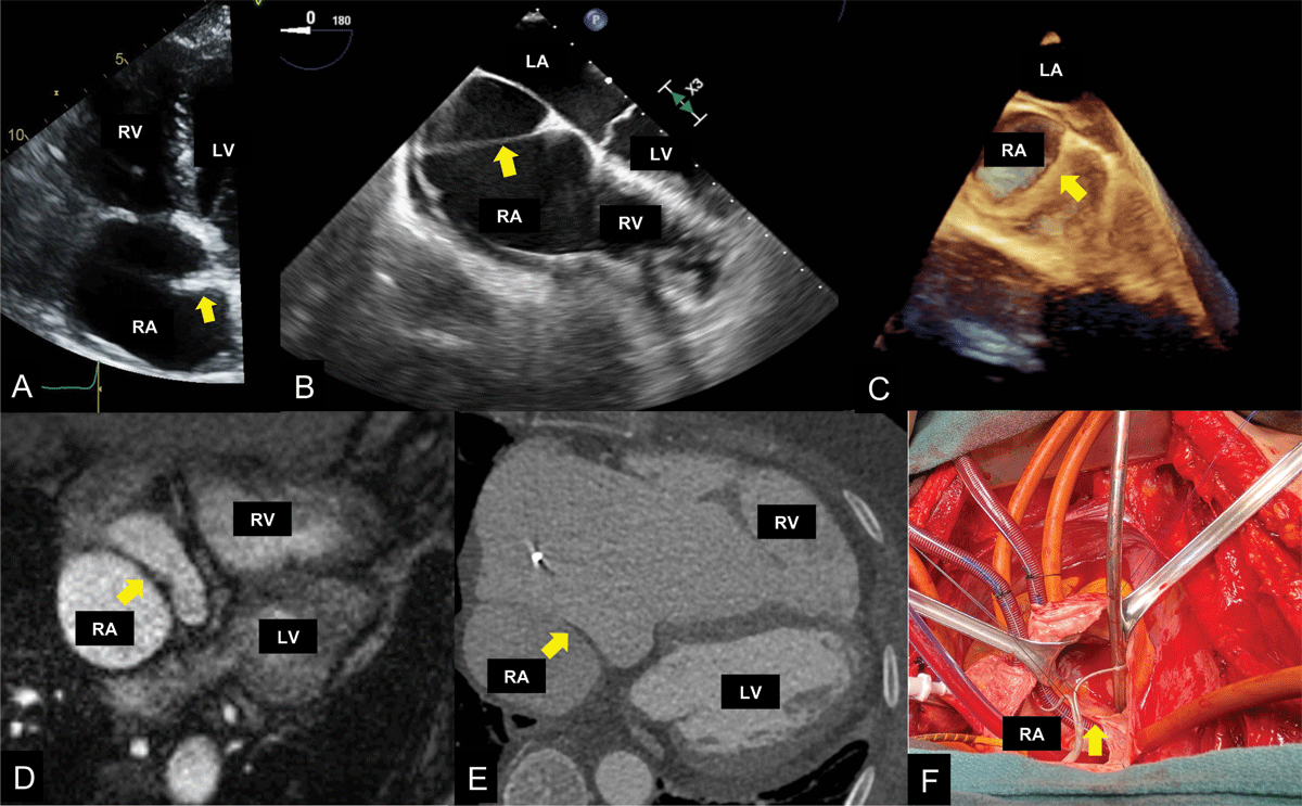
Figure 1
(A) Transesophageal echocardiogram (TEE) depicts a noncontinuous linear structure that seems to attach to the intra-atrial septum. (B) TEE further defines the membrane as a continuous structure, with color Doppler showing flow across membrane (see Video 1). (C) 3-dimensional (3D) TEE confirms what was seen in 2D imaging. (D) Cardiac magnetic resonance imaging confirms cor triatriatum dexter. (E) Coronary computed tomography angiography depicts low-attenuation structure traversing through right atrium. (F) Intraoperatively, cor triatriatum dexter was confirmed, but importantly, right ventricular inflow obstruction was excluded. RV: right ventricle; LV: left ventricle; LA: left atrium; RA: right atrium
Video 1
Transesophageal echocardiogram depicting continuous membrane. Color Doppler shows flow across this membrane. This also can be viewed at https://youtu.be/wfF14wMF4C4.