
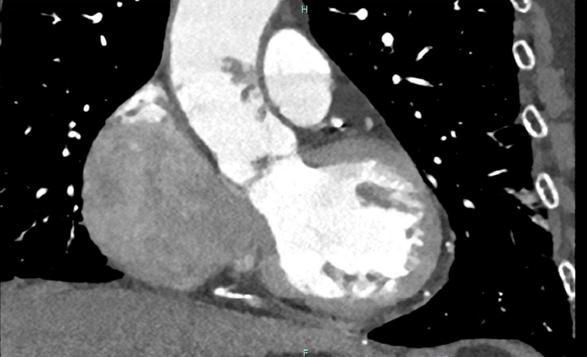
Figure 1
Longitudinal axial computed tomographic angiography image revealing a pedunculated aortic mass in the ascending aorta.
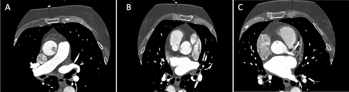
Figure 2
(A–C) Transverse axial images of coronary computed tomographic angiography at different axial levels demonstrate coronary contrast filling defects in the ascending aorta consistent with suspected aortic thrombus (finally determined to be primary aortic fibroelastoma by histopathology).
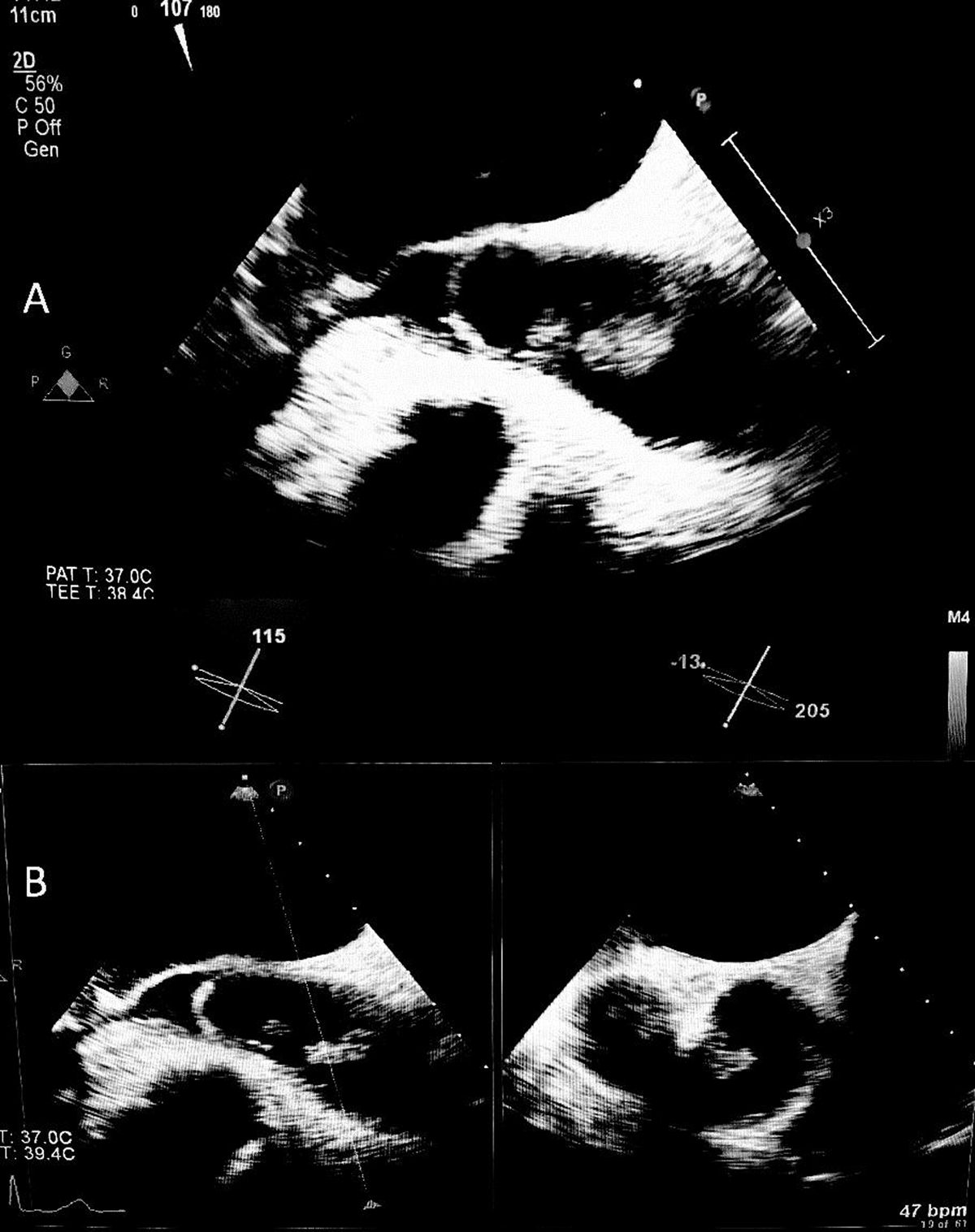
Figure 3
(A) Transesophageal view demonstrating a mobile pedunculated aortic mass in the ascending aorta, superior to the aortic valve. (B) The same aortic mass is visualized across an x-plane (perpendicular plane) view of the transesophageal echocardiogram.
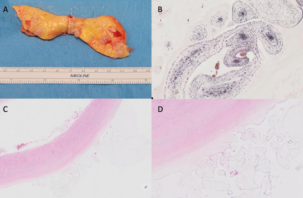
Figure 4
(A) Gross pathology of aortic fibroelastomas: the specimen presented is a part of the ascending aorta. The intimal surface demonstrates multiple papillary areas in different locations, with pink tan to yellowish discoloration with some mild focal calcification consistent with aortic fibroelastoma. (B) Verhoeff-Van Gieson/EVG stain highlights the elastic cores on histopathology images confirming the diagnosis of fibroelastoma. (C, D) Histopathology slides on different magnification scale demonstrate avascular fibroelastic tissue lined by a single layer of cells with multiple branching points, consistent with papillary fibroelastoma.