
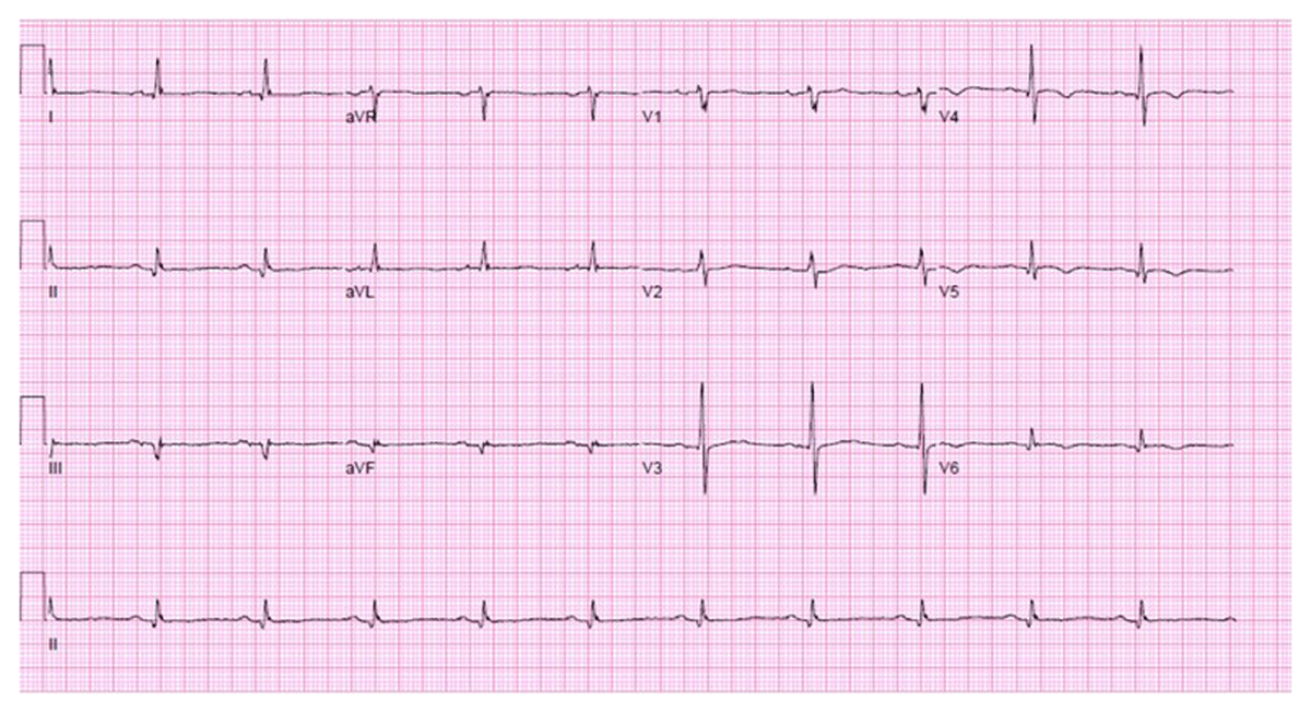
Figure 1
Electrocardiogram of patient presenting with exertional dyspnea and fatigue.
Question 1
Consider the options and find the answer in this video quiz, also at https://youtu.be/5BQTEHbMlew.
Video 1
Play the video to see what the transthoracic echocardiogram revealed, also at https://youtu.be/WHHURP04pNM.
Video 2
Play the video to see what the transthoracic echocardiogram revealed, also at https://youtu.be/n4PSr3e-18Q.
Video 3
Play the video to see what the transthoracic echocardiogram revealed, also at https://youtu.be/BHd8hmcOibY.
Question 2
Consider the options and find the answer in this video quiz, also at https://youtu.be/HYEAt-GqSPU.

Figure 2
Computed tomography showing a large bilobed posterolateral left ventricular pseudoaneurysm.
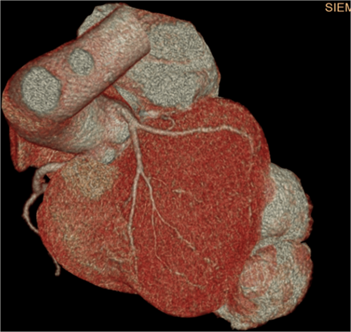
Figure 3
3-dimensional reconstructed computed tomography showing a large bilobed posterolateral left ventricular pseudoaneurysm.
Video 4
Computed tomography showing a large bilobed posterolateral left ventricular pseudoaneurysm, also at https://youtu.be/ks6FDJgg41g.
Question 3
Consider the options and find the answer in this video quiz, also at https://youtu.be/X4VrGOVJNbk.
Video 5
Cardiac fluorodeoxyglucose-positron emission tomography did not show uptake of fluorine-18-deoxyglucose in the pseudoaneurysm, also at https://youtu.be/_Yfb9SBNDYI.
Video 6
Cardiac magnetic resonance shows a basal–mid-lateral wall myocardial infarction with contained rupture resulting in a large (8.1 × 3.9 cm) left ventricular pseudoaneurysm, also at https://youtu.be/zcRz8J8gqxs.

Figure 4
Computed tomography done prior to the atrial fibrillation ablation did not show a pseudoaneurysm.
Video 7
Cardiac magnetic resonance 1 week post repair showed a thin myocardial repair graft at the mid-inferolateral segment of the left ventricle with mild biventricular dysfunction, also at https://youtu.be/AxCbNGwEZ6U.