
Figure 1
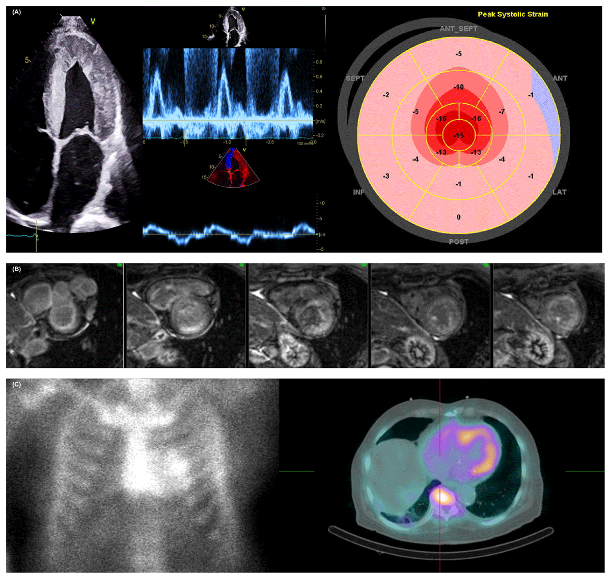
Multimodality imaging approach for cardiac amyloidosis (CA). (A) Increased left ventricular (LV) wall and interatrial thickness with sparkling texture are typically found in patients with CA (left). Evidence of restrictive LV filling, rapid E-wave deceleration time, high E/A ratio on mitral inflow Doppler, and low myocardial relaxation velocities on tissue Doppler imaging (middle). Corresponding “bullseye” map of the longitudinal strain pattern of the LV with a “cherry on top” sign (right). (B) Cardiac magnetic resonance imaging shows characteristic imaging of CA with diffuse and subendocardial late gadolinium enhancement. (C) 99mTc-PYP anterior planar (left) imaging and single-photon emission computed tomography/computer tomography (right) showing a Perugini Grade of 2 and 3, respectively.
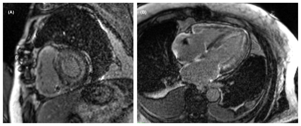
Figure 2
(A) Short-axis view and (B) long-axis view shows diffuse late gadolinium enhancement uptake (Seimens 1.5 Tesla).

Figure 3
T1 map (precontrast) shows the left ventricle with an elevation of native T1 values (Seimens 1.5 Tesla).

Figure 4
99mTc-PYP cardiac scintigraphy shows anterior planar imaging using heart-to-contralateral ratio semiquantitative scoring. Reproduced with permission from Springer Nature. doi: 10.1007/s10741-021-10174-x.
H/CL: heart to contralateral lung; ATTR: transthyretin amyloidosis
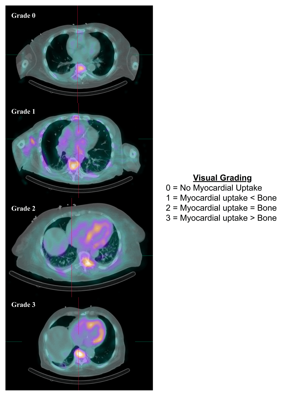
Figure 5
99mTc-PYP cardiac scintigraphy using single photo emission computer tomography/computed tomography visual scoring. Reproduced with permission from Springer Nature. doi: 10.1007/s10741-021-10174-x
Table 1
Reported non-transthyretin amyloidosis causes of false positive and false negative Tc-labeled cardiac scintigraphy. Reproduced with permission from Springer Nature, doi: 10.1007/s10741-021-10174-x
| FALSE POSITIVE | FALSE NEGATIVE |
|---|---|
| Amyloid light-chain amyloidosis | Early disease |
| Hydroxychloroquine toxicity | Delayed imaging |
| Rib fracture | Full length (type B, Phe64Leu, Ser77Tyr, and Thr59Lys) transthyretin fibrils |
| Pleural effusion | Small radiotracer injection dose |
| Valvular calcification | Short acquisition time |
| Blood pool | Pericardial effusion |
| Breast implants | Breast implants |
| Myocardial infarction (acute or subacute) | |
| Rare forms of cardiac amyloidosis | |
| APO-A1 mutation |