
Have a personal or library account? Click to login
A Case of Cardiac Arrest Caused by Air Embolism from Routine Root Canal Procedure Cover

A Case of Cardiac Arrest Caused by Air Embolism from Routine Root Canal Procedure

Open Access
|Sep 2022

Figures & Tables

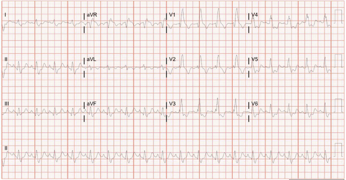
Figure 1

Electrocardiogram.
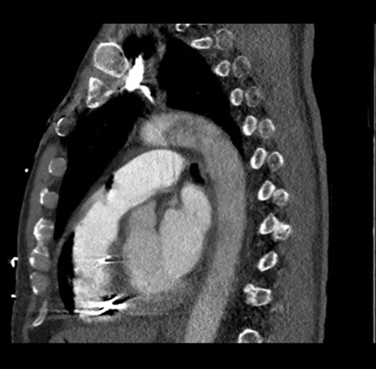
Figure 2

Computed tomography sagittal view demonstrating right ventricular air embolism with extension into the pulmonary trunk.
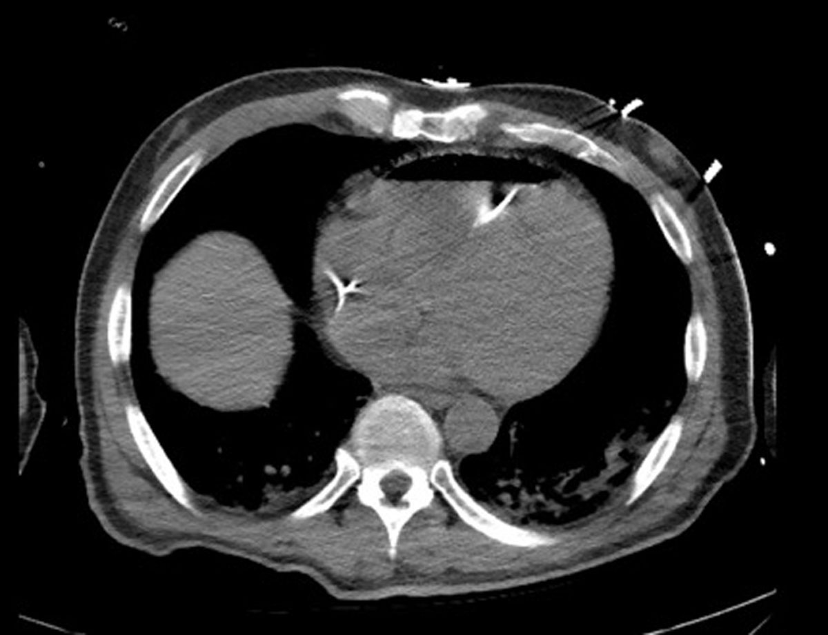
Figure 3

Computed tomography chest axial view showing presence of air in the right ventricle.
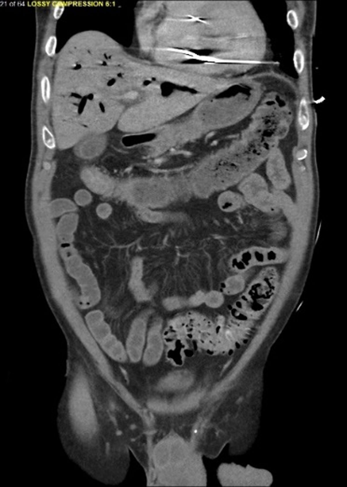
Figure 4

Computed tomography of the abdomen showing a venous air embolism in right ventricle and portal system.
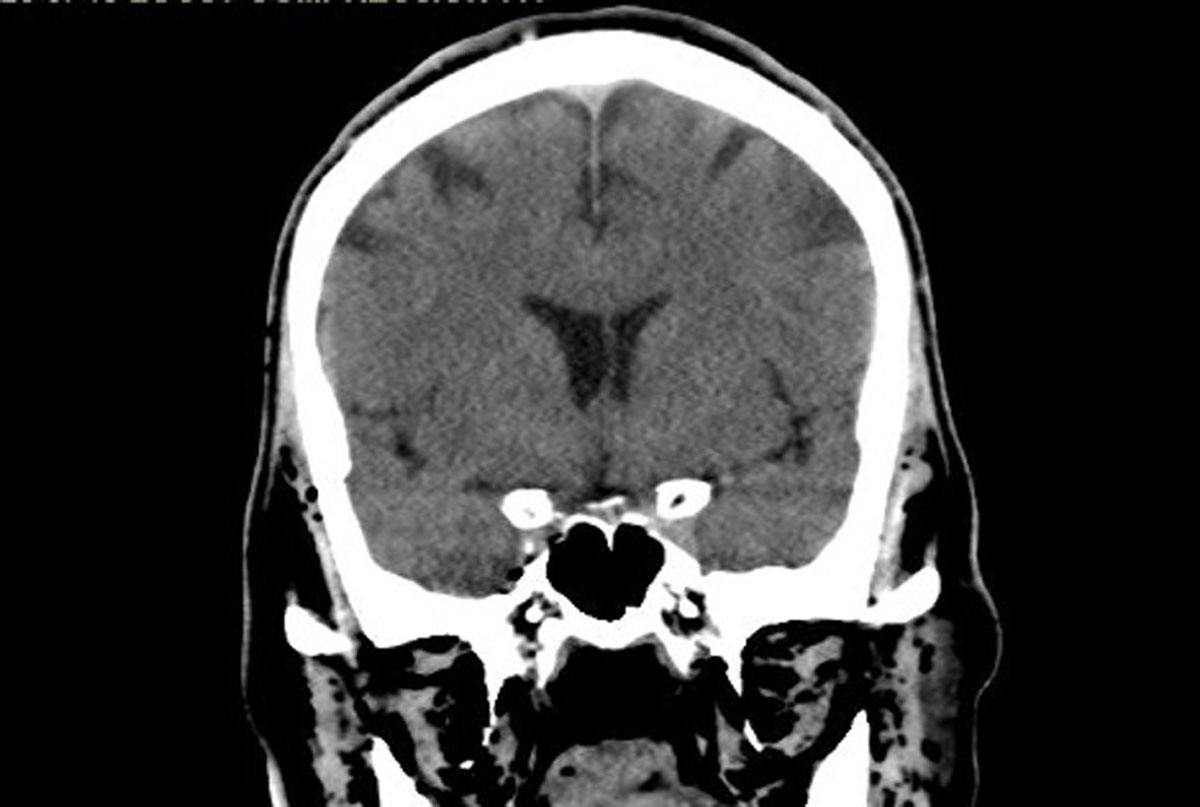
Figure 5

Computed tomography of the head revealing the presence of air in facial tissue.

DOI: https://doi.org/10.14797/mdcvj.1067 | Journal eISSN: 1947-6108
Language: English
Page range: 68 - 72
Submitted on: Nov 28, 2021
Accepted on: Jul 19, 2022
Published on: Sep 20, 2022
Published by: Houston Methodist DeBakey Heart & Vascular Center
In partnership with: Paradigm Publishing Services

© 2022 Akarsh Parekh, Joel McCormick, Atheer Hussain-Amin, Bryan Barnosky, Matthew Edwards, published by Houston Methodist DeBakey Heart & Vascular Center
This work is licensed under the Creative Commons Attribution-NonCommercial 4.0 License.