
Table 1
Differential diagnosis of chest pain in patients with COVID-19. ECG: electrocardiogram; STEMI: ST-elevation myocardial infarction; NSTEMI: non-ST-elevation myocardial infarction; ICA: invasive coronary angiogram; CAD: coronary artery disease; TTE: transthoracic echocardiogram; POCUS: point-of-care ultrasound; CCTA: coronary computed tomography angiography; JVP: jugular venous pressure; COVID-19: coronavirus disease 2019; PNA: pneumonia; CT: computed tomography; CTPA: computed tomography pulmonary angiography; LV: left ventricle; RWMA: regional wall motion abnormalities; MINOCA: myocardial infarction with nonobstructive coronary arteries; CMR: cardiac magnetic resonance imaging; LGE: late gadolinium enhancement; DVT: deep vein thrombosis; ACS: acute coronary syndrome
| DISEASE PROCESS | PRESENTATION | IMAGING FOR INITIAL AND SUBSEQUENT ASSESSMENT |
|---|---|---|
| Acute coronary syndrome | Ischemic chest pain with typical ECG changes and elevation of cardiac biomarkers |
|
| Cardiac tamponade | Dyspnea, tachypnea, elevated JVP, hypotension, pulsus paradoxus, electrical alternans on ECG |
|
| COVID-19 pneumonia | Cough, fever, myalgia, headache, dyspnea, loss of smell/taste, worsening hypoxemia |
|
| Heart failure, new onset, without hypotension | Progressive dyspnea, signs of congestion, S3 heart sound |
|
| MINOCA | Ischemic chest pain with ECG changes and modest elevation of cardiac biomarkers |
|
| Myocarditis | Variable but may include presentation of acute heart failure with new arrhythmias |
|
| Pericarditis | Pleuritic chest pain, diffuse ST elevations and PR depressions on ECG, pericardial friction rub |
|
| Pulmonary embolism | Dyspnea, pleuritic chest pain, signs and symptoms of DVT |
|
| Stress cardiomyopathy | Similar to ACS with signs of heart failure |
|
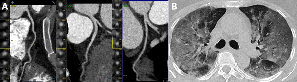
Figure 1
Coronary computed tomography (CT) angiography in a 34-year-old woman who presented with COVID-19 pneumonia. The patient had chest pain and minimal troponin elevation. (A) Coronaries were free of atherosclerosis or stenosis. (B) Chest CT shows advanced COVID-19 pneumonia.
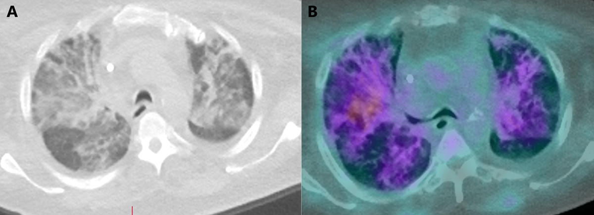
Figure 2
(A) Severe pulmonary infiltrates in a patient with recent COVID-19 infection referred for positive emission tomography myocardial perfusion imaging. (B) There is rubidium uptake in these pulmonary infiltrates.
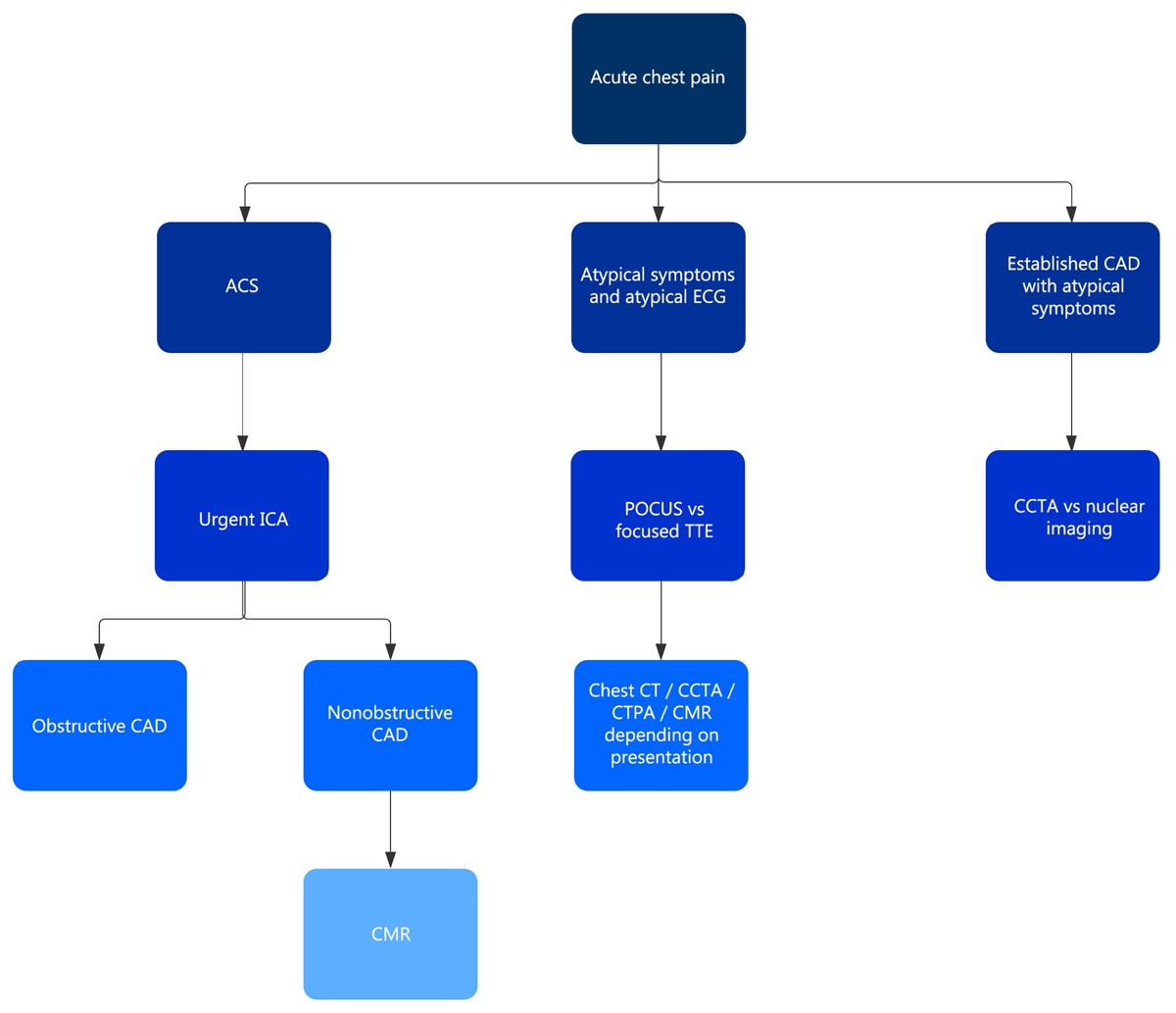
Figure 3
Flowchart for noninvasive imaging in patients with COVID-19 presenting with acute chest pain. ACS: acute coronary syndrome; ECG: electrocardiogram; CAD: coronary artery disease; ICA: invasive coronary angiography; POCUS: point-of-care ultrasound; TTE: transthoracic echocardiogram; CCTA: coronary computed tomography angiography; CT: computed tomography; CTPA: computed tomography pulmonary angiography; CMR: cardiac magnetic resonance imaging