
Have a personal or library account? Click to login
Renal Vein Thrombosis due to Posterior Nutcracker Syndrome Cover

Renal Vein Thrombosis due to Posterior Nutcracker Syndrome

Open Access
|Oct 2020

Figures & Tables

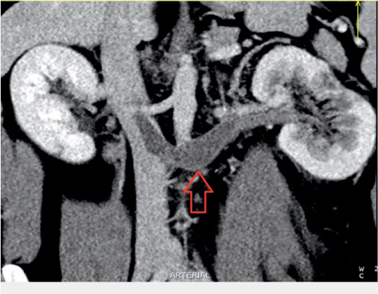
Figure 1.

Coronal computed tomography reformat showing thrombus-filled left renal vein extending into suprarenal inferior vena cava (arrow).
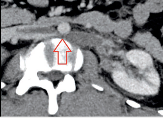
Figure 2.

Selected axial computed tomography indicating retroaortic course of thrombosed left renal vein with extrinsic compression by the abdominal aorta (arrow).
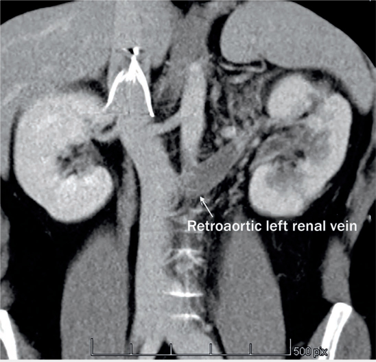
Figure 3.

Coronal computed tomography reformat showing suprarenal position of the inferior vena cava.

Language: English
Published on: Oct 1, 2020
Published by: Houston Methodist DeBakey Heart & Vascular Center
In partnership with: Paradigm Publishing Services

© 2020 Laura Lizama, Sergio Bran, Sven Kuestermann, Rudolf García-Gallont, published by Houston Methodist DeBakey Heart & Vascular Center
This work is licensed under the Creative Commons Attribution-NonCommercial 4.0 License.