
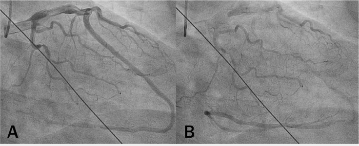
Figure 1.
Right anterior oblique (A) caudal and (B) distal views showing gradual opacification of left anterior descending artery as it turns around the apex, continues in the posterior interventricular groove, then makes another 180° turn at the crux to continue back to the apex.
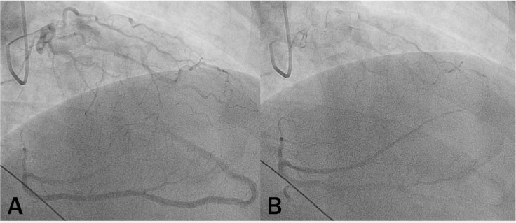
Figure 2.
Right anterior oblique (A) proximal and (B) distal cranial images allowing better visualization of distal left anterior descending artery running in the posterior interventricular groove, then turning around at the crux to continue back to the apex.
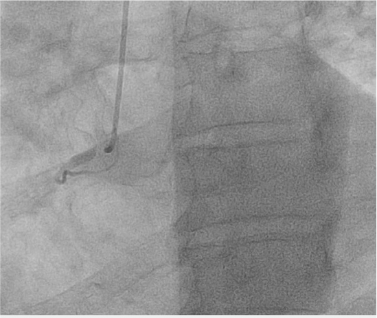
Figure 3.
Right coronary artery (RCA) in left anterior oblique (LAO) caudal plane; only rudimentary RCA is visualized in the LAO plane.
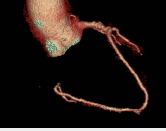
Figure 4.
Three-dimensional reconstruction of left anterior descending artery (LAD) on cardiac computed tomography angiography, which allows us to better appreciate the spatial orientation of the vessel. Unfortunately, the long course of the LAD did not allow adequate contrast passage to the most distal parts of the vessel.