
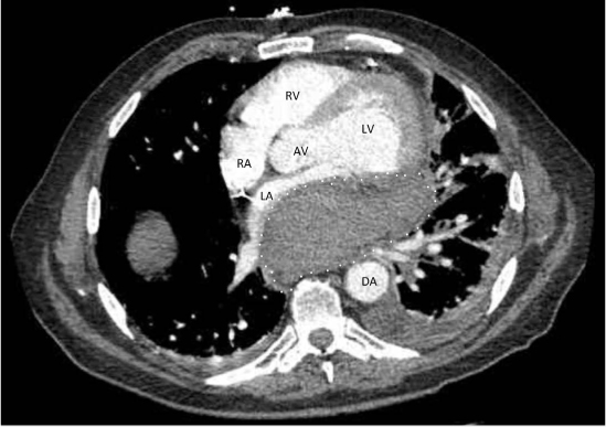
Figure 1.
Computer tomographic angiography image of the chest revealing a retrocardiac mass (white dotted line) measuring 6.1 × 9.5 × 8.7 cm effacing the left atrium and pulmonary veins. LV: left ventricle; RV: right ventricle; LA: left atrium; RA: right atrium; DA: descending thoracic aorta; AV: aortic valve
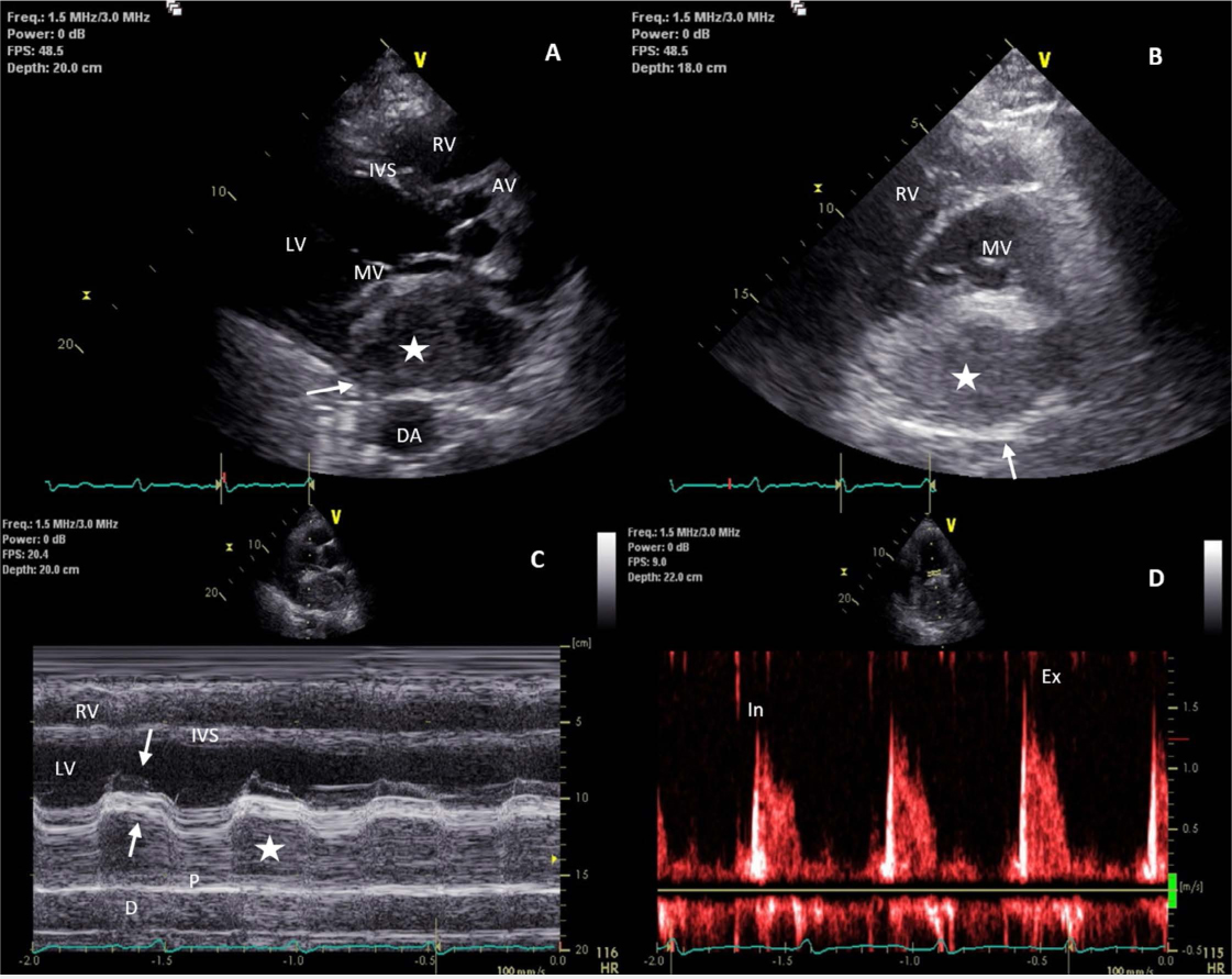
Figure 2.
Echocardiographic images of a large posteriorly located intrapericardial hematoma. (A) Parasternal long-access view showing severe compression of the left atrium by a posteriorly located echolucent mass (white star) contained within the pericardium (white arrow). The posterior leaflet of the mitral valve has been severely anteriorly displaced. (B) Parasternal short-access view depicts similar compression of the mitral valve with anteior displacement of the posterior leaflet of the mitral valve by the echolucent mass (white star). (C) M-mode image through the plane of the mitral valve. The image shows the posteriorly located echolucent mass (white star) as well as restricted motion of the posterior leaflet of the mitral valve (upgoing arrow) with normal motion of the anterior leaflet (downgoing arrow). (D) Continous wave Doppler through the mitral valve showing significant respiratory variation in mitral inflow velocities, with more than 30% variation from inspiration (In) to expiration (Ex) concerning for obstructed flow through the mitral valve. LV: left ventricle; RV: right ventricle; IVS: interventricular septum; MV: mitral valve; AV: aortic valve; DA: descending thoracic aorta; P: pericardium
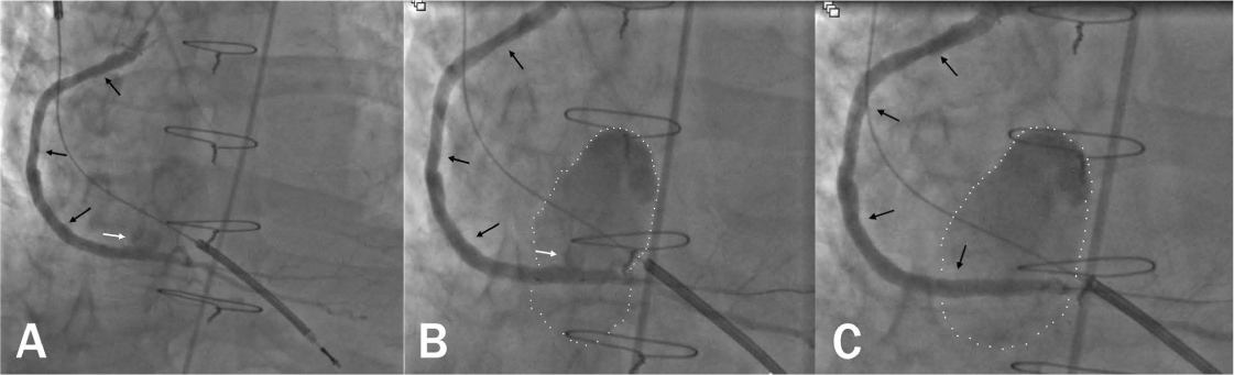
Figure 3.
Coronary bypass graft angiographic images of the saphenous vein graft (black arrows) to the right posterior descending coronary artery. (A) Angiographic image shows a perforation (white arrow) near the anastomosis of the bypass conduit and the native vessel. (B) Angiographic image post deployment of 2.8 × 16 and 3.5 × 15 mm Graftmaster RX covered stents (Abbott Vascular). Perforation is still noted (white arrow) after deployment of the stent, and a posteriorly located hematoma is also visualized (white dotted lines). (C) Angiographic image post stent deployment and successful balloon tamponade with a 4.0 mm NC Quantum Apex balloon (Boston Scientific). Posteriorly located hematoma is still present (white dotted line).
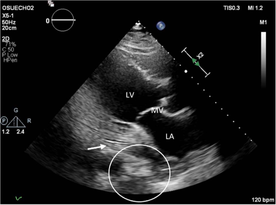
Figure 4.
Echocardiographic parasternal long-access image following video-assisted thoracic surgery to evacuate a large posteriorly located intrapericardial hematoma; also shows placement of a pericardial drain (white arrow) with relief of the compression on the left atrium and mitral valve. LV: left ventricle; LA: left atrium; MV: mitral valve