
Have a personal or library account? Click to login
Artificial Intelligence and Machine Learning in Cardiovascular Imaging Cover

Artificial Intelligence and Machine Learning in Cardiovascular Imaging

Open Access
|Oct 2020

Figures & Tables

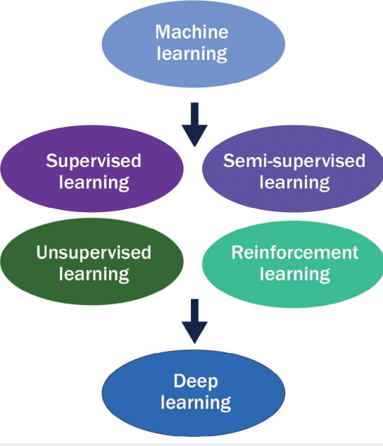
Figure 1.

Progression of machine learning.

Table 1.

Description of an array of studies that have leveraged machine learning applications in echocardiography, computed tomography angiography, nuclear imaging, and cardiac magnetic resonance imaging.20–22,28,29,31–34,37,38,41,43 CTA: computed tomography angiography; CAD: coronary artery disease; TPD: total perfusion deficit; MACE: major adverse cardiac events
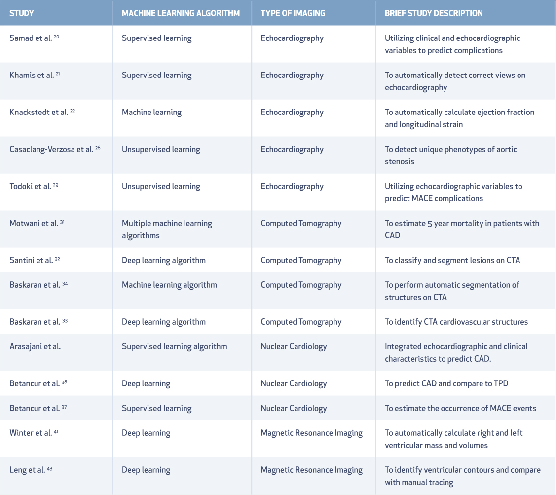
i1947-6094-16-4-263-t01.jpg
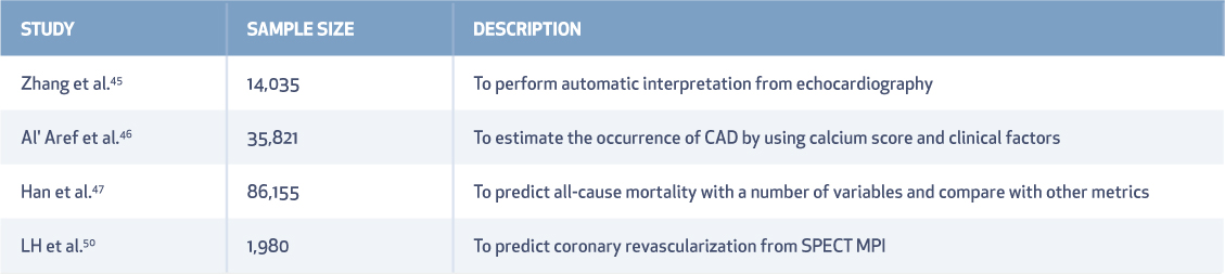
Table 2.

Summary of studies that have evaluated machine learning algorithms for prediction or automated interpretation.45–47,50 SPECT: single photon emission computed tomography; MPI: myocardial perfusion imaging; CAD: coronary artery disease

i1947-6094-16-4-263-t02.jpg
Language: English
Page range: 263 - 271
Published on: Oct 1, 2020
Published by: Houston Methodist DeBakey Heart & Vascular Center
In partnership with: Paradigm Publishing Services

© 2020 Karthik Seetharam, James K. Min, published by Houston Methodist DeBakey Heart & Vascular Center
This work is licensed under the Creative Commons Attribution-NonCommercial 4.0 License.