
Have a personal or library account? Click to login
Large Sinus of Valsalva Aneurysm Complicated by Thrombus Formation Cover

Large Sinus of Valsalva Aneurysm Complicated by Thrombus Formation

Open Access
|Jul 2020

Figures & Tables

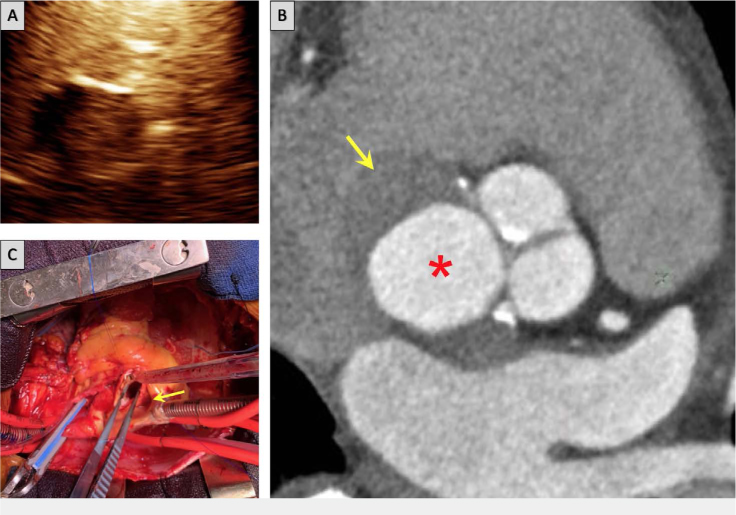
Figure 1.

(A) Transthoracic echocardiogram shows a filling defect within the noncoronary cusp after administration of ultrasound contrast, suggestive of a thrombus. (B) Computed tomography coronary angiogram shows aneurysmal noncoronary sinus (*indicates dilated noncoronary cusp aneurysm, arrow indicates thrombus). (C) Intraoperative image shows aortic root at the level of sinus of Valsalva and the noncoronary cusp aneurysm (arrow).

Language: English
Published on: Jul 1, 2020
Published by: Houston Methodist DeBakey Heart & Vascular Center
In partnership with: Paradigm Publishing Services

© 2020 Milan Ravishankar, Ali Ghergherehchi, Jing Liu, Ernesto Jimenez, Umair Khalid, published by Houston Methodist DeBakey Heart & Vascular Center
This work is licensed under the Creative Commons Attribution-NonCommercial 4.0 License.