
Table 1.
Multimodality imaging in prosthetic valve dysfunction by prosthetic valve position.
TEE: transesophageal echocardiography; MDCT: multidetector computed tomography; CMR: cardiovascular magnetic resonance imaging; PVL: paravalvular leak
| TYPE OF PROSTHETIC VALVE DYSFUNCTION | 3D TEE | MDCT | CMR |
|---|---|---|---|
| Obstruction (pannus vs thrombus) | |||
| Mitral valve | +++ | +++ | ++# |
| Aortic valve | ++* | +++ | ++# |
| Tricuspid/pulmonic valve | ++ | ++ | ++# |
| Regurgitation (valvular vs PVL) | |||
| Mitral valve | +++ | ++± | +++ |
| Aortic valve | ++* | ++± | +++ |
| Tricuspid/pulmonic valve | ++ | ++± | +++ |
| Endocarditis (vegetation, abscess, fistula, pseudoaneurysm) | |||
| Mitral valve | +++ | ++** | ++** |
| Aortic valve | ++* | ++** | ++** |
| Tricuspid/pulmonic valve | ++* | ++** | ++** |
| + fair, ++ good, +++ excellent |
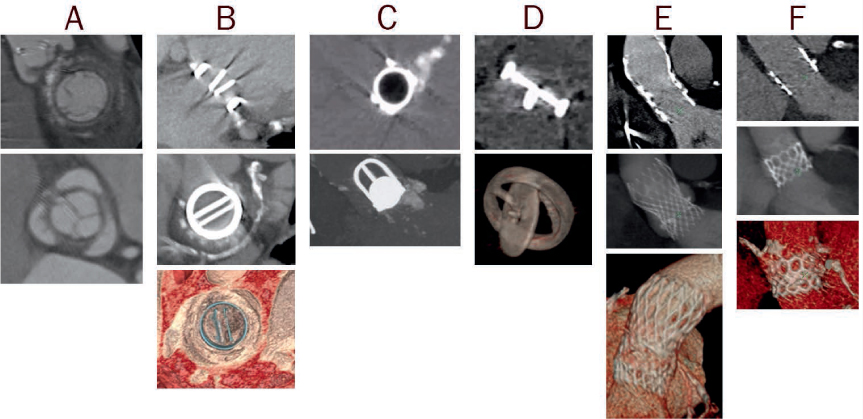
Figure 1.
Examples of computer tomography images of prosthetic valves, including (A) biologic stented, (B) mechanical bileaflet tilting disc, (C) mechanical ball-in-cage, (D) mechanical single tilting disc, (E) transcatheter self-expandable, and (F) transcatheter balloon-expandable.
Table 2.
Differentiation of etiology of prosthetic valve obstruction by pannus versus thrombus.
*timeframe from surgery; MV: mitral valve; AV: aortic valve; TV: tricuspid valve; INR: International Normalized Ratio; AF: atrial fibrillation/flutter; HU: Hounsfield units
| PANNUS | THROMBUS | |
|---|---|---|
| Timeframe* | ⩾ 12 mos, commonly ⩾ 5 yrs | Occurs at any timeframe (often mixed with pannus at later stages) |
| Valve location and type | MV > AV Mechanical = bioprosthetic | TV > MV = AV Mechanical > bioprosthetic |
| Morphology | Fixed, small dense mass composed of fibrin material Bright echodensity Usually involve suture lines and subprosthetic location | Mobile mass (usually larger than pannus) Soft echodensity Any location (usually supravalvular) |
| Clinical characteristics | ||
| INR | No effect | Increased risk with low INR (increased risk with AF and low cardiac output) |
| Symptoms | Insidious symptoms | Rapidly progressive symptoms, higher risk of stroke |
| Response to anticoagulation | No or minimal response | ⩾ 50% decrease in prosthesis gradient |
| CT HU attenuation | HU ⩾ 145 | HU ⩾ 90 |
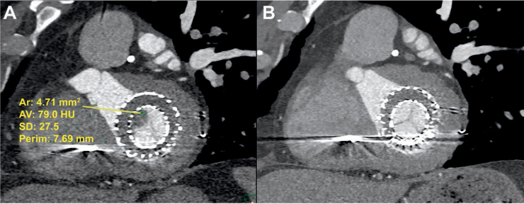
Figure 2.
Transcatheter mitral valve prosthesis (A) with thrombus (HU < 90) noted on multidetector computed tomography and (B) with resolution of thrombus after anticoagulation on repeat computed tomography after 5 months. HU: Hounsfield units
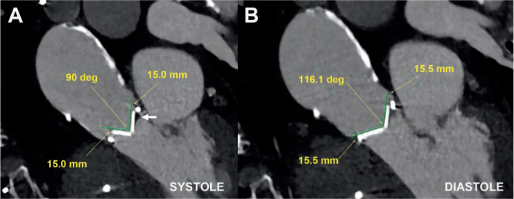
Figure 3.
Multidetector computed tomography demonstrating (A) abnormal opening angle of 90° in systole and (B) normal closure in diastole in a patient with severe mechanical prosthetic aortic stenosis due to mixed pannus and thrombus (arrow).
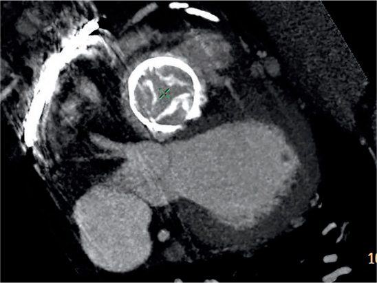
Figure 4.
Degenerated bioprosthetic tricuspid valve with malcoaptation of calcified leaflets in systole resulting in severe tricuspid regurgitation.
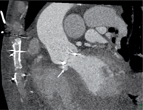
Figure 5.
Prosthetic valve endocarditis complicated by prosthetic valve partial dehiscence and fistula between the aorta and left ventricle cavity (arrow).
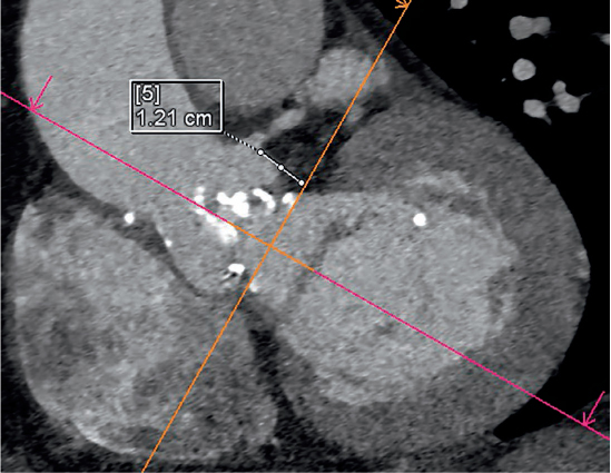
Figure 6.
Multidetector computed tomography demonstrating measurement of the left coronary ostial height of 12 mm for planned aortic valve-in-valve procedure.
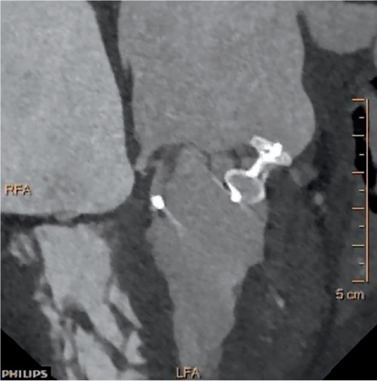
Figure 7.
Multidetector computed tomography reconstruction for paravalvular leak (PVL) with subsequent PVL closure.
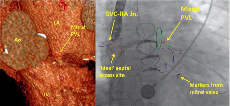
Figure 8.
Multidetector computed tomography fusion imaging for planning of paravalvular leak closure. Ao: aorta; LV: left ventricle: LA: left atrium; PVL: paravalvular leak; SVC-RA Jn.: superior vena cava-right atrial junction
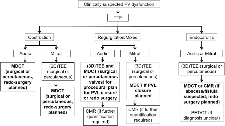
Figure 9.
Role of multidetector computed tomography (MDCT) for management of patients with prosthetic valve dysfunction. TTE: transthoracic echocardiography; TEE: transesophageal echocardiography; CMR: cardiac magnetic resonance imaging; PVL: paravalvular leak; PET/CT: positron emission tomography/computed tomography