
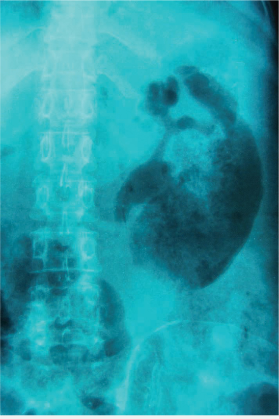
Figure 1.
Plain film of the abdomen shows significant amount of gas in the left kidney.
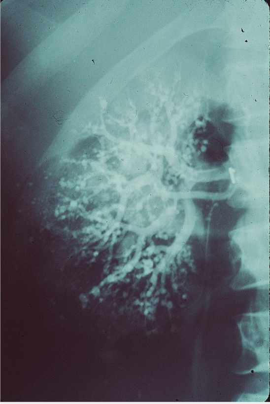
Figure 2.
Renal arteriogram shows macroaneurysms of the renal circulation.
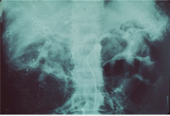
Figure 3.
Abdominal arteriogram shows macroaneurysms of the mesenteric circulation.
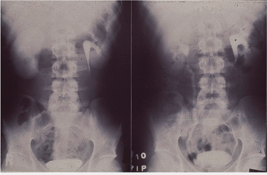
Figure 4.
(Left) Initial intravenous pyelogram (IVP) shows a dense nephrogram of the right kidney with no visualization of the collecting system compared to the contralateral kidney. (Right) Repeat IVP shows a normal study with full visualization of the normal-appearing collecting system bilaterally.
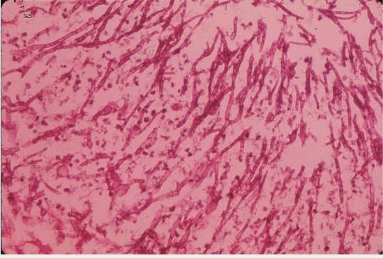
Figure 5.
Spontaneously voided tissue sample shows septated hyphae and mycelia (Aspergillus fumigatus).
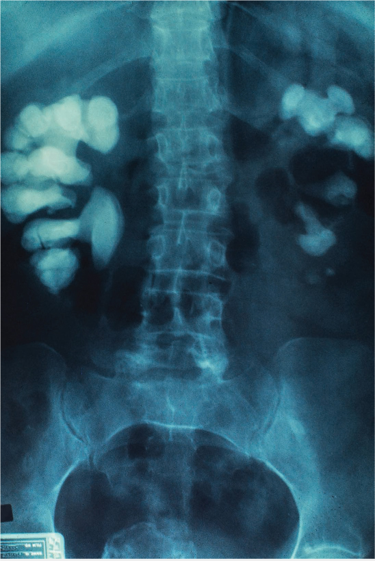
Figure 6.
Plain film of the abdomen shows bilateral large radiopaque stones consistent with stag horn calculi.