
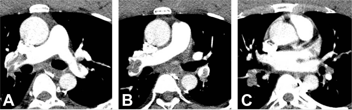
Figure 1.
Computed tomography angiography of chest showing (A) bilateral proximal, (B) segmental, and (C) subsegmental pulmonary embolisms.
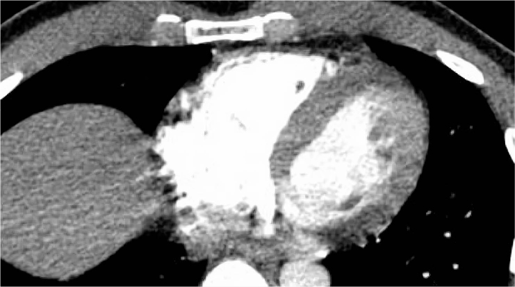
Figure 2.
Computed tomography angiography of the chest showing right-to-left ventricular size ratio of approximately 0.9 concerning for early signs of right heart strain.
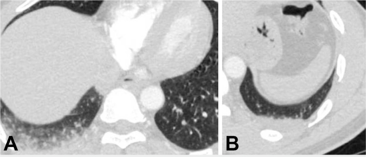
Figure 3.
Computed tomography angiography of chest showing bilateral ground glass opacities concerning for an atypical infectious process.