
Table 1.
SPECT versus PET radiotracers and technical characteristics.
SPECT: single photon emission computed tomography; PET: positron emission tomography; KeV: kiloelectron volts
| CHARACTERISTIC | SPECT | PET |
|---|---|---|
| Radiotracer | Generator | Generator/cyclotron |
| Energy | 80–140 KeV | 511 KeV |
| Attenuation | More | Less |
| Half-life | 6–72 hours | 1.5–13 minutes |
| Radiation | Greater | Significantly less |
| Study duration | 3–4 hours | 30–40 minutes |
| Stress | Exercise > pharmacological | Pharmacological only in clinical use |
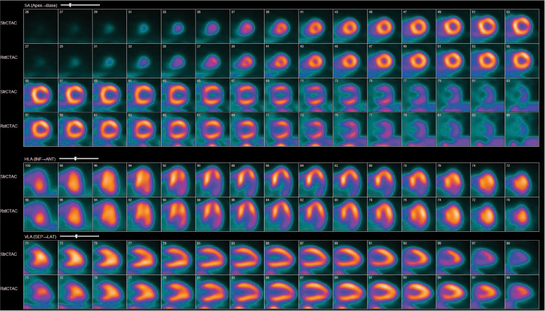
Figure 1.
Positron emission tomography 82rubidium scan depicting moderate myocardial ischemia in the lateral wall. Rows 1,3,5,7 were obtained at peak stress, whereas rows 2,4,6,8 show the corresponding rest images. SA: short axis; HLA: horizontal long axis; VLA: vertical long axis
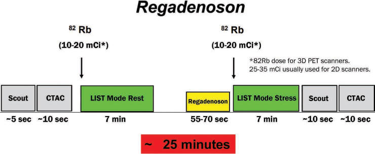
Figure 2.
Currently employed PET MPI pharmacological (regadenoson) stress protocol. The use of 3-dimensional scanners equipped with digital detectors has allowed for the lowering of delivered 82rubidium dose down to between 10 and 20 mCi. PET: positron emission tomography; MPI: myocardial perfusion imaging; 82 Rb: 82rubidium; CTAC: computed tomography attenuation correction; CAC: coronary artery calcium
Table 2.
Characteristics of available positron emission tomography radiotracers.
Reproduced with permission from Al Badarin et al.13
| CHARACTERISTIC | 82RUBIDIUM | 13N-AMMONIA | 15O-WATER |
|---|---|---|---|
| Supplied | Generator | Cyclotron | Cyclotron |
| Half-life | 76 s | 9:96 min | 2.09 min |
| Uptake mechanism | Active extraction | Active extraction | Freely diffusible |
| Positron range in water | 1.6 mm | 0.28 mm | 0.5 mm |
| Image quality | Very good | Excellent | Uninterpretable |
| Radiotracer uptake characteristics | Adequate | Very good | Excellent |
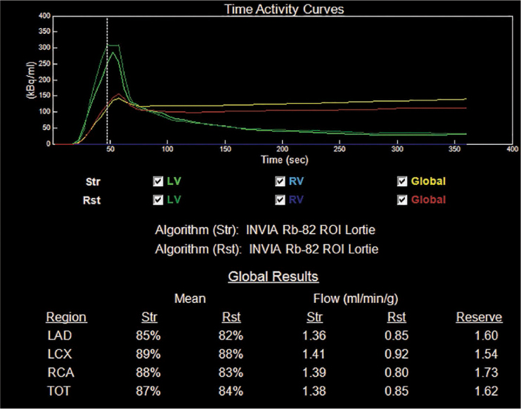
Figure 3.
Time-activity curves for the assessment of myocardial blood flow in a patient with nonobstructive coronary artery disease on coronary angiogram. The global coronary flow reserve is reduced, indicating microvascular dysfunction. Several models are available for coronary flow reserve estimation, but the 1-tissue compartment model depicted in this figure is the most widely employed. Str: stress; Rst: rest; LV: left ventricle; RV: right ventricle; LAD: left anterior descending artery; LCX: left circumflex artery; RCA: right coronary artery; TOT: total