
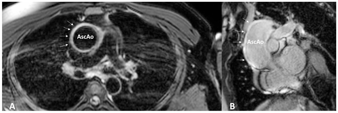
Figure 1.
(A) Axial cardiac magnetic resonance slice demonstrating diffuse, circumferential ascending aortic wall thickening (arrows) on T2-weighted fat saturation imaging. (B) Late gadolinium enhancement imaging reveals hyperenhancement (arrows) of the ascending aorta and proximal aortic arch. AscAo: ascending aorta
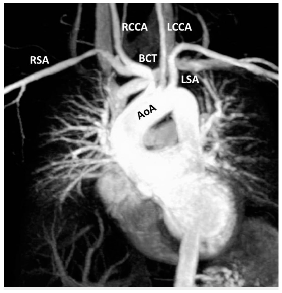
Figure 2.
Three-dimensional gadolinium-enhanced maximum intensity projection rendering of aortic arch and major branch vessels. No significant stenoses, occlusions, or aneurysms are seen. AoA: aortic arch; RSA: right subclavian artery; LSA: left subclavian artery; RCCA: right common carotid artery; LCCA: left common carotid artery; BCT: brachiocephalic trunk
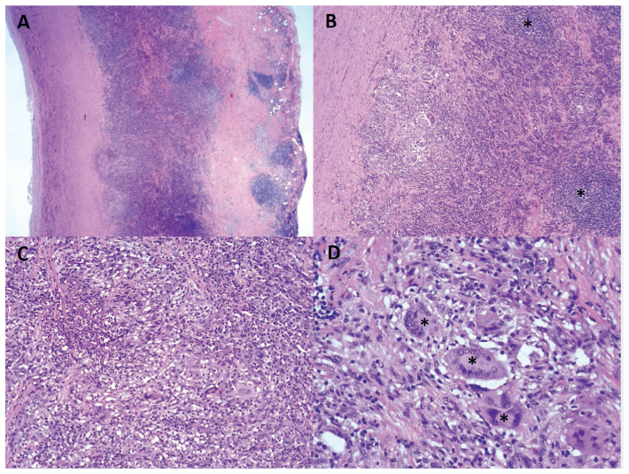
Figure 3.
(A) Histology of the ascending aortic wall demonstrating abnormal wall thickness up to 0.7 cm, (B, asterisks) lymphoid aggregates in the tunica media and adventitia, (C) predominantly lymphoplasmacytic inflammatory cell infiltrates, and (D, asterisks) multinucleated giant cells.