
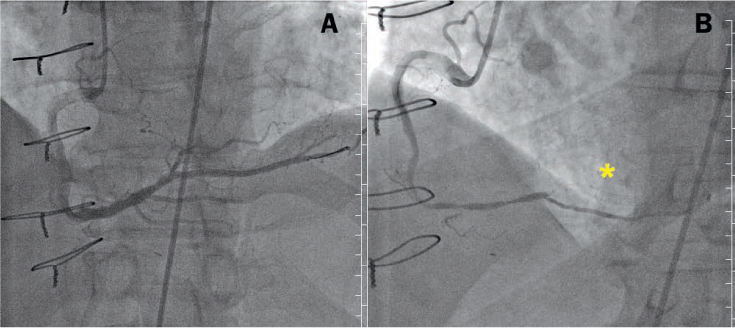
Figure 1.
Coronary angiograms in right anterior oblique projections demonstrate (A) successful perfusion of a culprit right posterolateral branch artery immediately after primary percutaneous coronary intervention, and (B) occlusion of the right coronary artery shortly thereafter. The coronary stent is visible (asterisk).
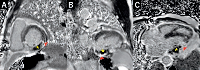
Figure 2.
Phase-sensitive inversion recovery cardiac magnetic resonance applied to late gadolinium enhancement in (A) short axis midpapillary, (B) 2-chamber, and (C) 3-chamber views demonstrate hypoenhanced inferior and inferolateral microvascular obstruction zones (asterisks) surrounded by hyperenhanced infarct (arrowheads).