
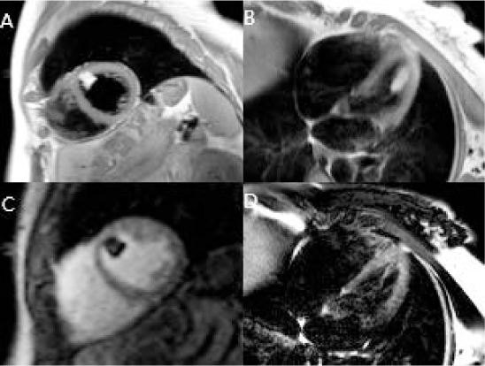
Figure 1.
Cardiac magnetic resonance imaging showing hyperintense signal of the left ventricular mass on dark blood T1 weighted imaging in (A) short axis and (B) 4-chamber long axis views, (C) lack of perfusion of the mass with first-pass administration of gadolinium-based contrast, and (D) complete signal void with T2 fat suppression imaging.
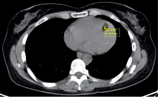
Figure 2.
Axial computed tomography slice showing low Hounsfield units of −82 when the left ventricular mass is sampled, consistent with adipose tissue.
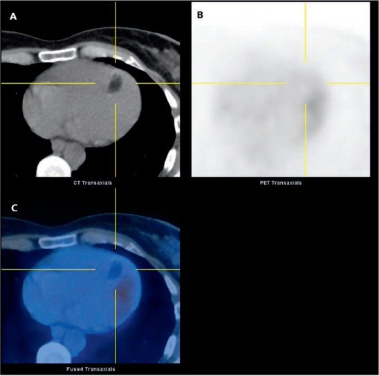
Figure 3.
Combined positron emission (PET) and computed tomography (CT) images showing (A) a hypodense left ventricular mass with virtually no uptake of fluorodeoxyglucose (18F-FDG), suggesting (B) a benign etiology due to lack of metabolic activity. (C) A hybrid PET/CT axial slice.