
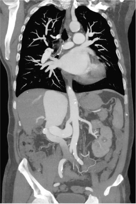
Figure 1.
Initial computed tomography angiography shows a missing right kidney, enlarged remnant of the right renal artery with s-shaped course, and a huge communication to the inferior vena cava that contrasted intensely during the arterial phase due to a high-flow left-to-right shunt.
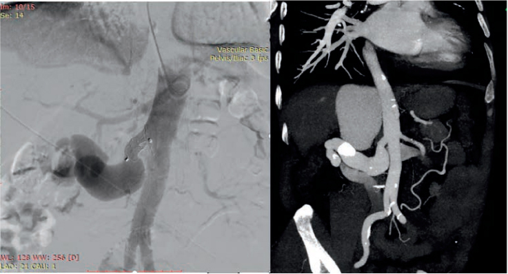
Figure 2.
Left: The final ostial position of the Occlutech PDA Occluder (Occlutech International) with the inferior vena cava contrasted later in relation to the arterial phase. Right: Control computed tomography, which was performed on the next day.
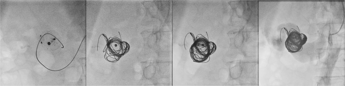
Figure 3.
Stepwise embolization with coils of the arteriovenous communication using the stable position of the patent ductus arteriosus/left atrial appendage occluders for an anchoring maneuver to avoid distal coil migration. Note the attenuation of the contrast intensity in the inferior vena cava.
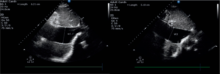
Figure 4.
Initial (left) and 1-month follow-up (right) echocardiograms show pronounced regression of the inferior vena cava maximal diameter from 62.1 mm to 51 mm.
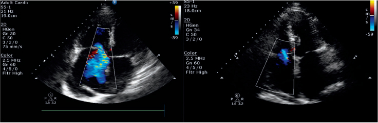
Figure 5.
Initial (left) and 1-month follow-up (right) echocardiograms show an impressive reduction of the third-degree tricuspid regurgitation to “trace” and reduction of the right atrium.
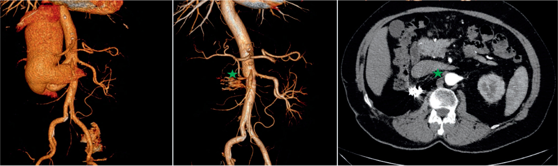
Figure 6.
Comparison between the initial (left) and 24-month follow-up (center) computed tomography angiography and follow-up computed tomography (right) shows stable position of the embolization materials, complete isolation of the aortocaval communication, and full interruption of the left-to-right shunt. The green star marks the inferior vena cava.