
A 30-year-old woman with a history of neonatal repair of infracardiac obstructed total anomalous pulmonary venous return (TAPVR) presented with dyspnea on exertion. Cardiac magnetic resonance imaging and computed tomography performed at presentation showed an anomalous connection—an unligated vertical vein—between the pulmonary venous confluence and portal venous system (Figure 1 A, B). The right ventricle was enlarged with depressed function. Transseptal catheterization was performed, and left lower pulmonary venography showed opacification of the confluence with return into the left atrium and into the portal vein through the vertical vein. An Agilis steerable sheath (Abbott Vascular) permitted optimal cannulation of the vertical vein, which measured 16.6× 14 mm (Figure 1 C). A 22-mm AMPLATZER Vascular Plug II (Abbott Vascular) was deployed through a 7-Fr Pinnacle Destination sheath (Terumo Medical) (Figure 1 D). After deployment, there was no pressure gradient between the left lower pulmonary vein and the left atrium. The patient reported improved exercise tolerance during follow-up.
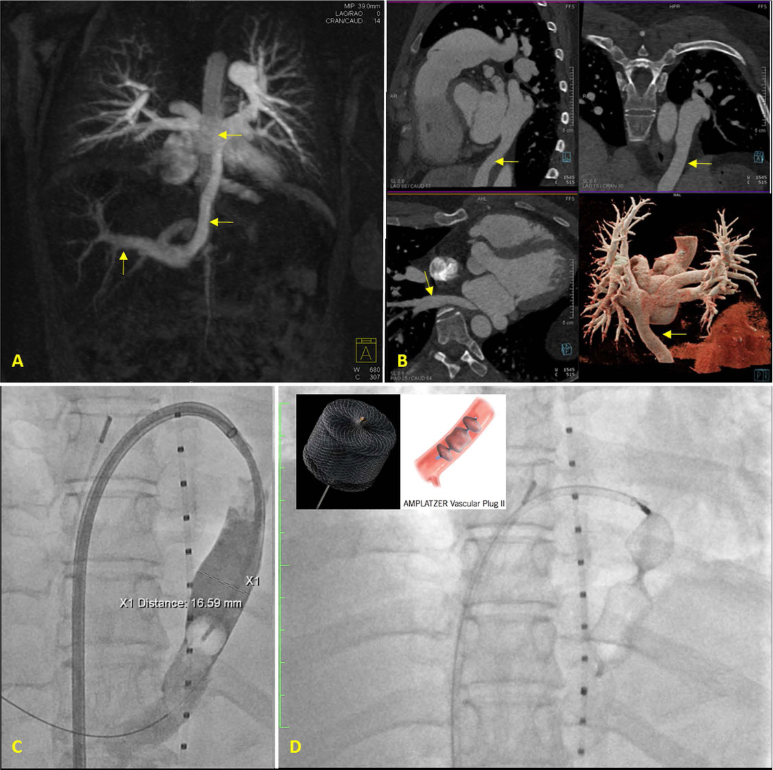
Figure 1.
(A) Cardiac magnetic resonance imaging showing an anomalous connection, the vertical vein, between the pulmonary venous confluence and the hepatic portal venous system. (B) Cardiac computed tomography demonstrating the vertical vein. (C) The vertical vein measured 16.6 × 14 mm in size. (D) 22-mm AMPLATZER Vascular Plug II in place was deployed in the vertical vein.
In TAPVR, the anomalous pulmonary veins return to a venous confluence, which then returns to the systemic venous circulation via a single “vertical vein.” In some cases, the vertical vein drains into the hepatic venous circulation, thus creating a left-to-right shunt that leads to right-sided volume overload. Some operators favor leaving the vertical vein unligated, particularly in obstructed TAPVR, while most favor ligation to remove the source of left-to-right shunt.1,2 This case depicts the successful transcatheter embolization of a persistent vertical vein.
Notes
[1] Conflict of Interest Disclosure: Dr. Chinnadurai is a full-time senior staff scientist at Siemens Healthcare, USA. Dr. Lin is a data monitoring committee member of ACI Clinical and speaker for Abiomed.