
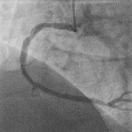
Figure 1.
Diagnostic angiography showing proximal right coronary artery lesion (left anterior oblique 25/cranial 10).
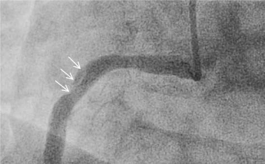
Figure 2.
Magnified view of proximal right coronary artery lesion (white arrows, left anterior oblique 25/cranial 10).
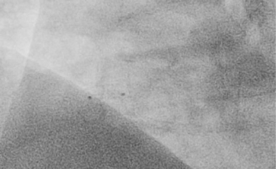
Figure 3.
Fluoroscopic still frame showing withdrawal of all equipment except retained delivery balloon (black fluoroscopic markers, left anterior oblique 30/cranial 10).
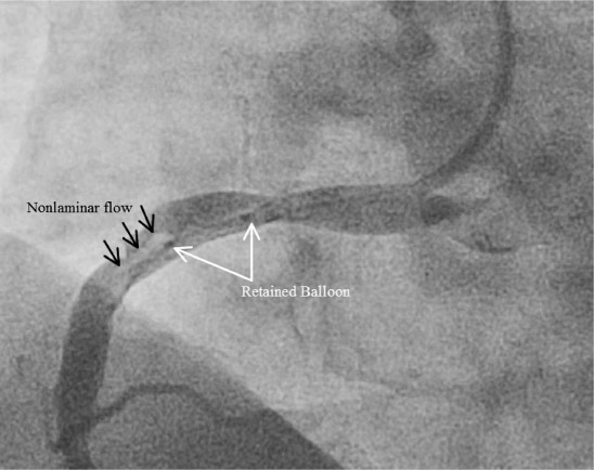
Figure 4.
Angiographic view after right coronary artery re-engagement showing retained balloon (white arrows), nonlaminar blood flow (black arrows, left anterior oblique 30/cranial 10).
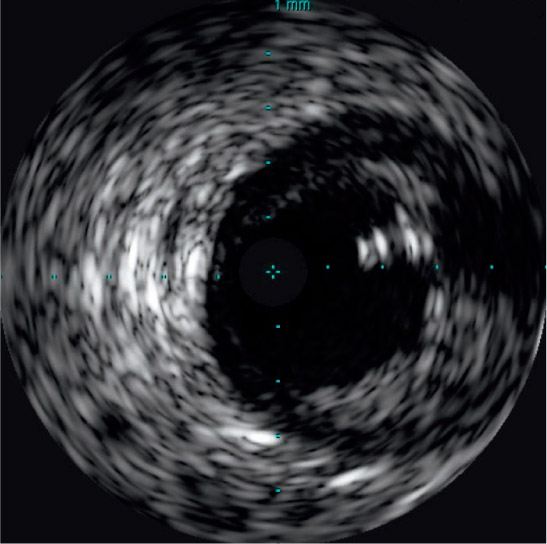
Figure 5.
Intravascular ultrasound demonstrating retained wire (2 to 3 o'clock position).
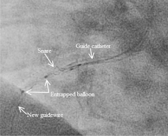
Figure 6.
Angiographic view of retrieval equipment during snare attempt (left anterior oblique 25/cranial 10).
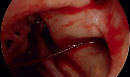
Figure 7.
Intraoperative view of proximal aorta and right coronary artery ostium (3 o'clock position) with retained wire fragment.
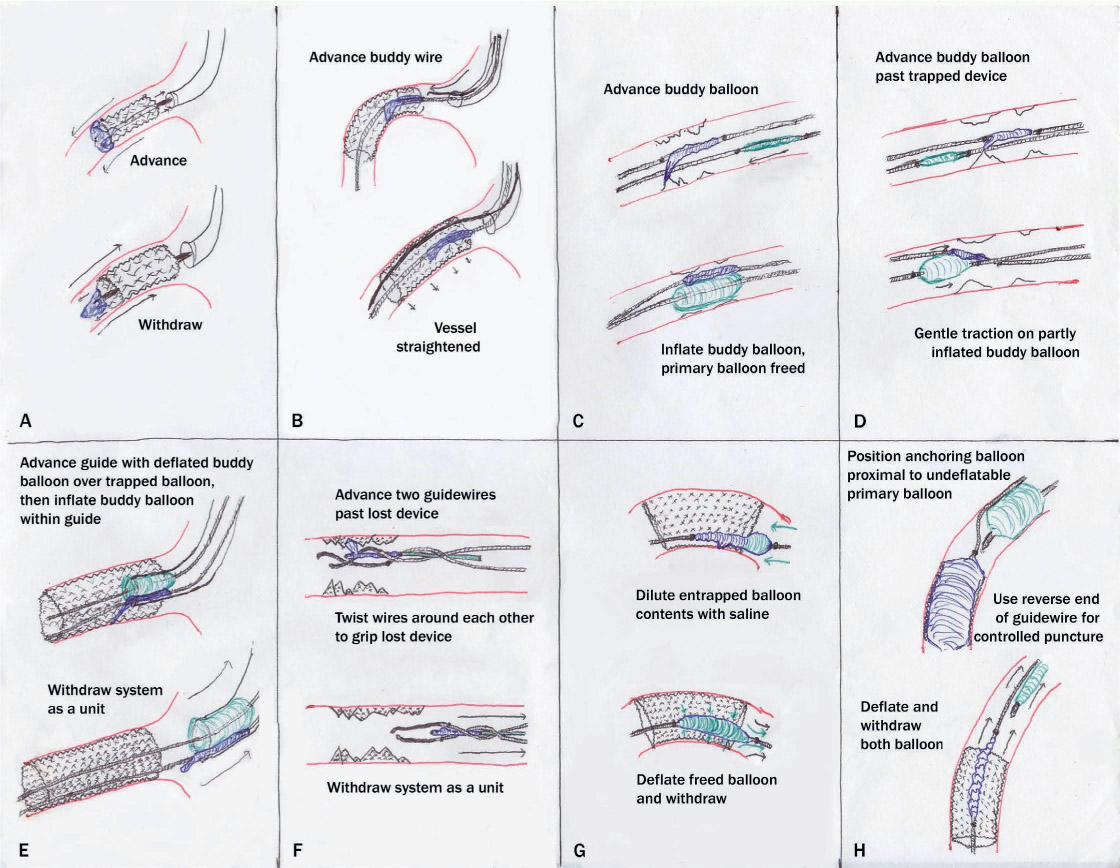
Figure 8.
Diagrammatic representation of possible percutaneous methods of device retrieval. (A) Advancement of entrapped balloon until free, followed by withdrawal. (B) Use of stiffer “buddy” wire to straighten segment containing trapped balloon and release balloon. (C) Advancement and inflation of a buddy balloon to free entrapped balloon. (D) Inflation of buddy balloon distal to entrapped device followed by gentle traction. (E) Entrapped balloon anchored inside guide catheter by anchoring balloon followed by withdrawal. (F) Two-wire technique, with guidewires entwined around entrapped device followed by withdrawal. (G) Attempted dilution of entrapped balloon contents with saline to free device. (H) Controlled puncture of incompletely deflated balloon by reverse guidewire and anchoring balloon.