
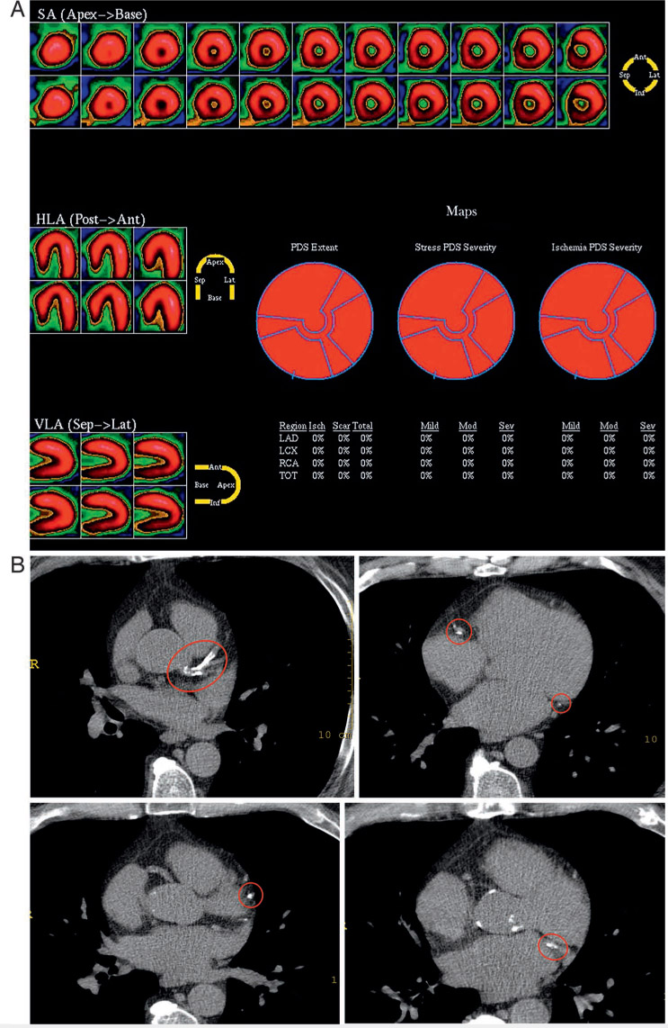
Figure 1.
A 65-year-old man with a history of hypertension, hyperlipidemia, cigarette smoking, and recent onset atypical chest pain. Exercise treadmill test results showed exercise duration of 8.5 minutes with a maximal heart rate of 125 bpm and no symptoms or ST changes. Duke treadmill score was 8.5 (low risk). (A) Stress myocardial perfusion images were normal. (B) Despite a normal functional test, the coronary artery calcium score (CACS) was severe at 740, indicating high risk for subsequent cardiac events. Red circles highlight areas of coronary calcification. SA: short axis; HLA; horizontal long axis; VLA: vertical long access; PDS: perfusion defect size; LAD: left anterior descending; LCX: left circumflex; RCA: right coronary artery; TOT: total.