
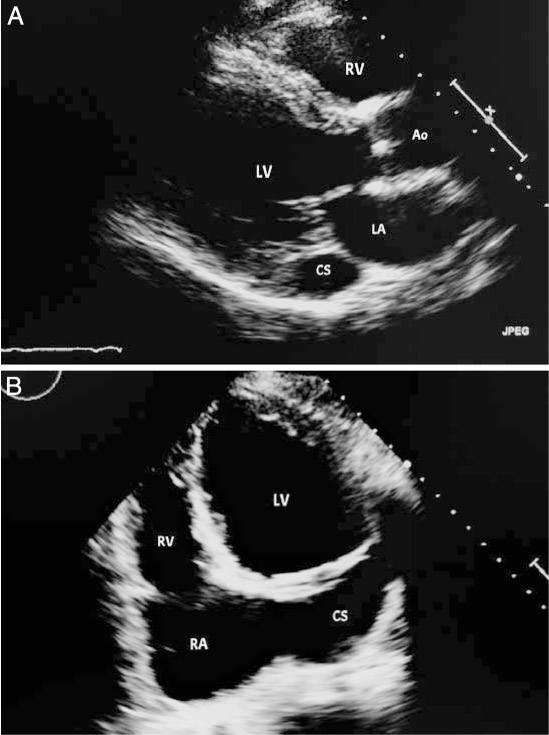
Figure 1.
(A) Two-dimensional transthoracic echocardiogram with a parasternal long view showing dilated coronary sinus, raising the suspicion for persistent left superior vena cava (PLSVC). (B) Apical view showing dilated coronary sinus, also raising the suspicion for PLSVC. LV: left ventricle; LA: left atrium; RA: right atrium; CS: coronary sinus; RV: right ventricle; Ao: aorta
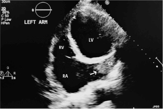
Figure 2.
Two-dimensional transthoracic echocardiogram with an apical view and injection of saline contrast through an IV catheter in the left upper extremity demonstrates saline contrast entering the coronary sinus (arrow) prior to the right atrium and right ventricle, confirming persistent left superior vena cava. RA: right atrium; RV: right ventricle; LV: left ventricle; CS: coronary sinus
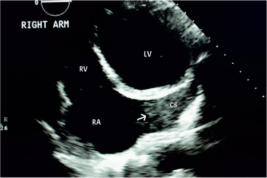
Figure 3.
Two-dimensional transthoracic echocardiogram with an apical view and injection of saline contrast through an IV catheter in the right arm demonstrates opacification of the coronary sinus (arrow) prior to the right atrium and right ventricle, confirming the absence of right superior vena cava. RA: right atrium; RV: right ventricle; LV: left ventricle; CS: coronary sinus
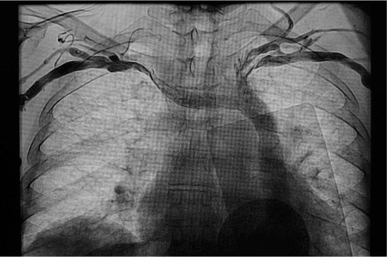
Figure 4.
Venogram with injections from left and right subclavian veins confirms persistent left superior vena cava and absent right superior vena cava (RSVC) with the right subclavian vein draining into the left SVC.