
Have a personal or library account? Click to login
Primary Nonbacterial Thrombotic Endocarditis Presenting with Bowel Infarction Secondary to Superior Mesenteric Artery Embolism Cover

Primary Nonbacterial Thrombotic Endocarditis Presenting with Bowel Infarction Secondary to Superior Mesenteric Artery Embolism

Open Access
|Jul 2018

Figures & Tables

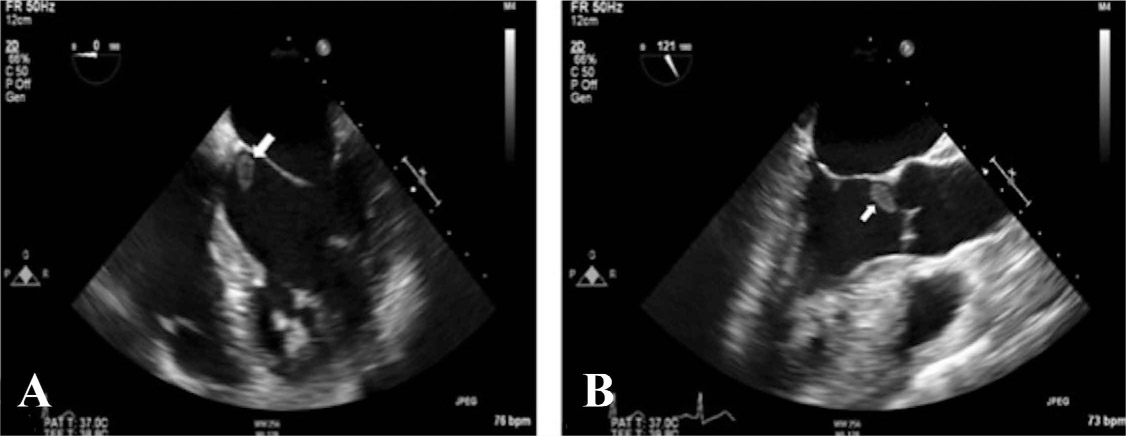
Figure 1.

Transesophageal echocardiogram. (A) Mid-esophageal (ME) apical 4-chamber view, and (B) ME aortic valve long-axis view showing a pedunculated mass (arrows) attached to interannular fibrosa of aortic valve.
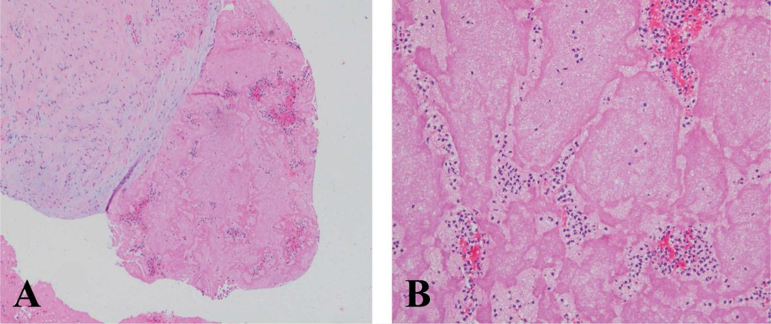
Figure 2.

(A) Histopathology of valve lesion consisted predominantly of homogeneous thrombotic material staining bright pink in routine hematoxylin and eosin stain (right); upper left shows small fragment of free edge of valve with early reactive fibrosis and lack of acute inflammation. (B) Higher power of thrombotic material that comprised most of the lesion shows typical pattern of acute thrombus; no evidence of infective organisms was seen. Original magnification: × 40 (A); × 100 (B).

Language: English
Page range: 228 - 231
Published on: Jul 1, 2018
Published by: Houston Methodist DeBakey Heart & Vascular Center
In partnership with: Paradigm Publishing Services

© 2018 Eduardo A. Rodriguez, Muhammad W. Choudhry, Paul J. Boor, Patrick T. Roughneen, Tareq Abu Sharifeh, published by Houston Methodist DeBakey Heart & Vascular Center
This work is licensed under the Creative Commons Attribution-NonCommercial 4.0 License.