
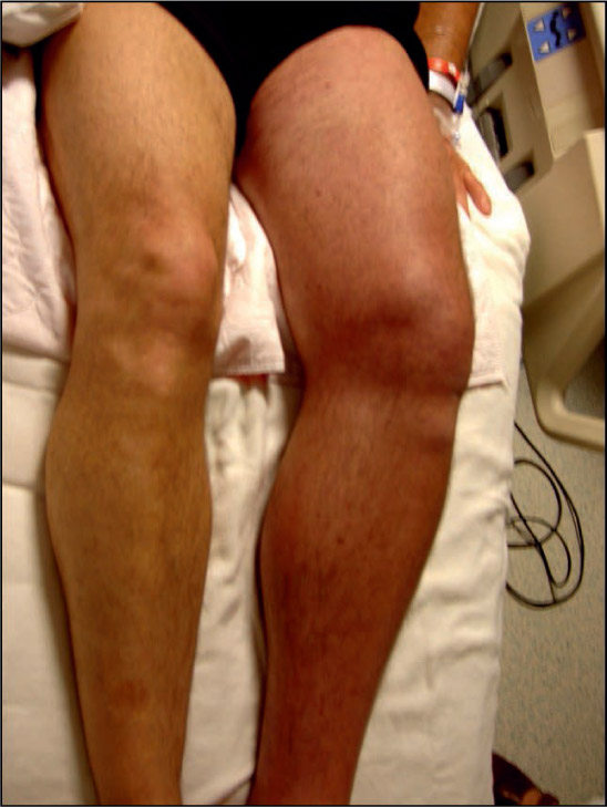
Figure 1.
Photograph of swollen, painful, discolored left leg (phlegmasia cerulea dolens) in a man with extensive iliofemoral DVT. The patient had received 5 days of anticoagulation therapy with low-molecular-weight heparin.
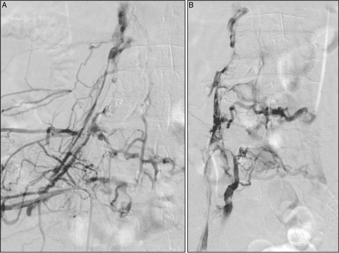
Figure 2.
An ascending diagnostic venogram performed via the ipsilateral popliteal vein in the prone position demonstrates extensive iliofemoral venous occlusion, shown in both (A) anteroposterior (AP) and (B) oblique views. Note the superimposed thrombus in the AP view. The patient was treated with pharmacomechanical catheter-directed thrombolysis.
Table 1.
Signs and symptoms of post-thrombotic syndrome (PTS) comprising the Villalta Score. Rank the severity of each symptom and sign on a scale of 0 (none) to 3 (severe), then add them up to determine a patient's Villalta Score for PTS.
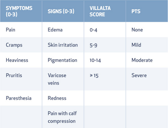
Table 2.
Primary outcomes of the ATTRACT trial. PDCT: pharmacomechanical catheter-directed thrombolysis; PTS: post-thrombotic syndrome
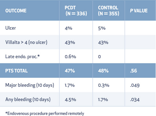
Table 3.
Secondary outcomes of the ATTRACT trial. PDCT: pharmacomechanical catheter-directed thrombolysis; PTS: post-thrombotic syndrome; VTE: venous thromboembolism
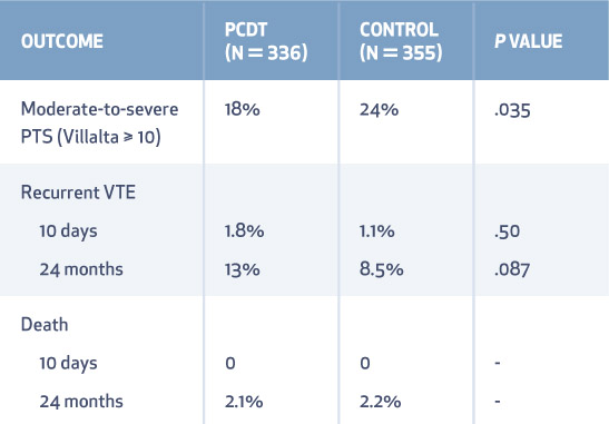
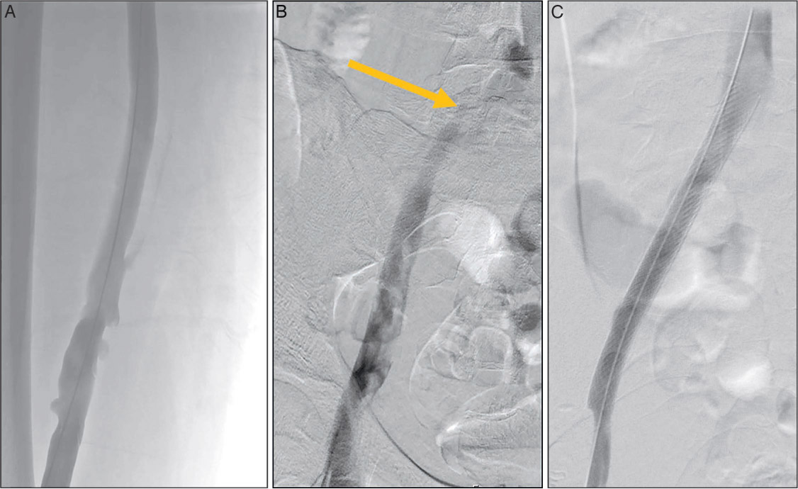
Figure 3.
Venogram in the prone position after pharmacomechanical catheter-directed thrombolysis shows (A) patency of the iliofemoral venous system. (B) There is a persistent stenosis of the left common iliac vein due to compression by the right common iliac artery (arrow). (C) After stenting of the residual common iliac vein stenosis, there is unobstructed venous drainage into the vena cava.
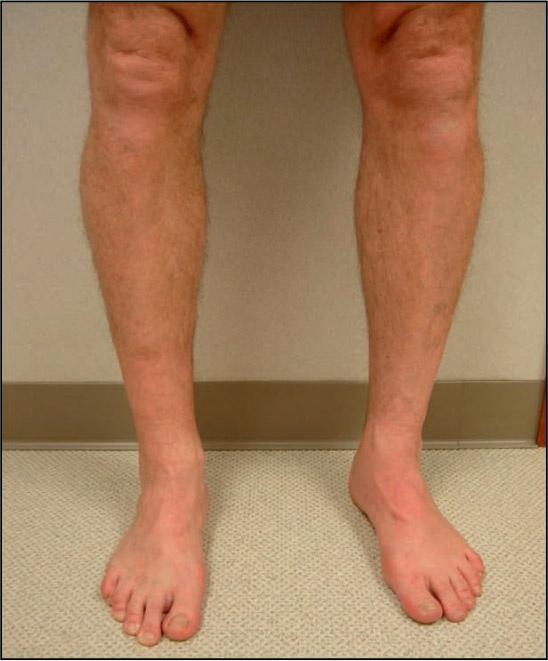
Figure 4.
At 12-month follow-up, the patient is asymptomatic with patent veins, no edema, and normal venous valve function.
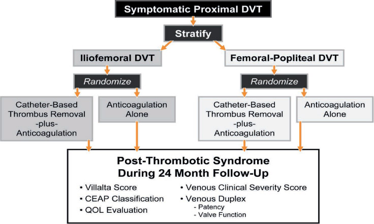
Figure 5.
Stratification and randomization sequence and outcome measures for the ATTRACT Trial. DVT: deep vein thrombosis; CEAP: a classification system to standardize the reporting and treatment of chronic venous disorders based on clinical, etiological, anatomical, and pathophysiological manifestations; QOL: quality of life