
Have a personal or library account? Click to login
Endovascular Therapy for Central Venous Thrombosis Cover

Endovascular Therapy for Central Venous Thrombosis

Open Access
|Jul 2018

Figures & Tables

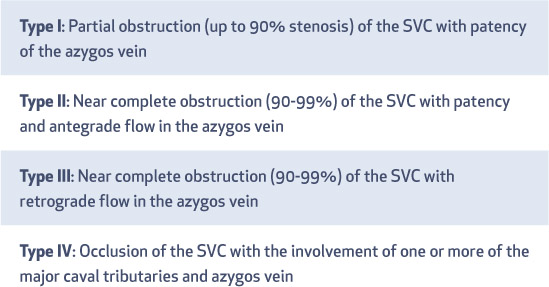
Table 1.

Stanford and Doty venographic classification of superior vena cava (SVC) obstruction.

i1947-6094-14-3-214-t01.jpg
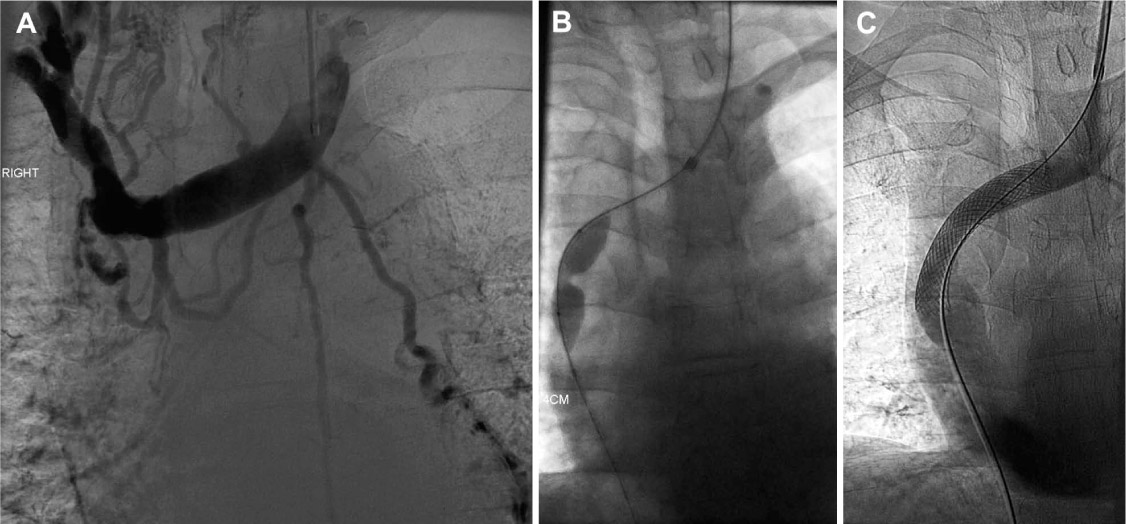
Figure 1.

A 63-year-old man with superior vena cava reconstruction with the WALLSTENT (Boston Scientific Corp.) following obstruction secondary to a hemodialysis line. (A) Pre-stent venogram, (B) pre-stent dilatation with balloon, and (C) post-stent venogram.
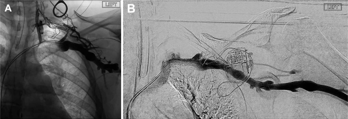
Figure 2.

A 34-year-old woman with left arm deep vein thrombosis secondary to cardiac pacemaker insertion for Brugada syndrome. The procedure included removal and replacement of the cardiac pacemaker, first rib resection, and insertion of a nitinol venous stent (Cook Zilver Vena). (A) Pre-stent venogram and (B) post-stent venogram.

Language: English
Page range: 214 - 218
Published on: Jul 1, 2018
Published by: Houston Methodist DeBakey Heart & Vascular Center
In partnership with: Paradigm Publishing Services

© 2018 Adam M. Gwozdz, Justinas Silickas, Alberto Smith, Prakash Saha, Stephen A. Black, published by Houston Methodist DeBakey Heart & Vascular Center
This work is licensed under the Creative Commons Attribution-NonCommercial 4.0 License.