
Figure 1.
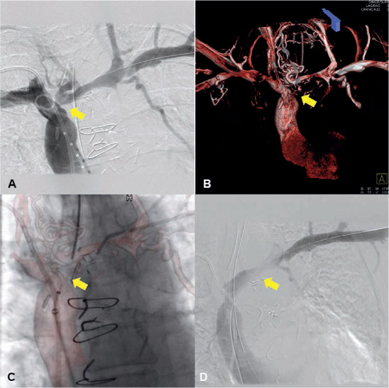
Intraoperative cone-beam CT imaging of a 63-year-old female with facial puffiness and left upper extremity swelling. (A) A venogram revealed occlusion of the left innominate vein. (B) Intraoperative cone-beam CT with contrast injection showed the 3D anatomy of venous collaterals and the occluded portion of the native innominate vein as a narrow stub (yellow arrow). (C) The entire 3D volume-rendered cone-beam CT reconstruction was overlaid onto real-time fluoroscopy for image guidance during venous recanalization attempts. (D) A venogram after recanalization documenting contrast flow across the left innominate vein. CT: computed tomography
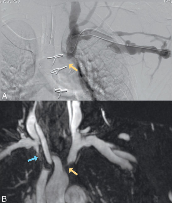
Figure 2.
(A) Digital subtraction angiography images acquired after iodinated contrast injection in left subclavian vein demonstrated occluded left innominate vein (yellow arrow). (B) Coronal maximum-intensity projection rendering of subtracted magnetic resonance venography images acquired after blood pool contrast agent injection (Feraheme) demonstrated perivascular information around the occluded right (blue arrow) and left innominate veins (yellow arrow) that were not visualized by luminal venography.
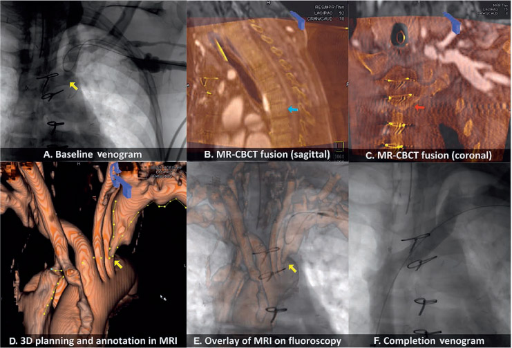
Figure 3.
Illustration of MRI-cone-beam CT image fusion and overlay on fluoroscopy for guidance during recanalization on a 55-year-old female with central venous occlusion after failed prior recanalization attempts. (A) A baseline venogram demonstrated complete occlusion of the left innominate vein (yellow arrow). (B, C) Intraoperative noncontrast cone-beam CT was acquired and coregistered with MRI dataset (T1-VIBE) using the spine (blue arrow) and sternum (red arrow) as alignment landmarks. (D) T1-VIBE and subtracted MR images demonstrated a venous lesion at the aortic arch level, where the path of the vein was electronically annotated. (E) This annotated vessel path was then overlaid on 2D fluoroscopy during the recanalization attempts. Accuracy of overlay was checked using wires/catheters at multiple fluoroscopic projections without the need for an additional venogram. (F) A completion venogram showed recanalized left innominate vein after stenting. MRI: magnetic resonance imaging; CT: computed tomography; CBCT: cone beam computed tomography