
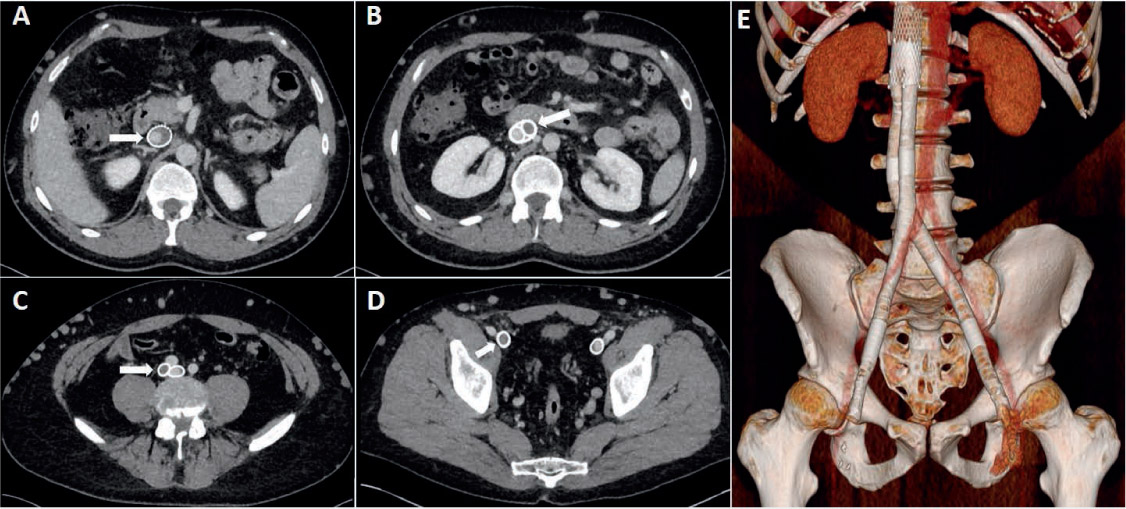
Figure 1.
Acute in-stent thrombosis. A filling defect can be seen in the inferior vena cava (IVC) stent (A), common iliac vein stent (B, C), and external iliac vein stent (D). (E) Three-dimensional reconstruction of the venous system demonstrating stenting from the IVC superiorly down to the common femoral vein inferiorly. This patient was successfully treated with catheter-directed thrombolysis.
Table 1.
Molecular imaging studies of venous thrombi using computed tomography (CT) and magnetic resonance imaging (MRI) techniques. FDG: fluorodeoxyglucose; PET: positron emission tomography; Gd: gadolinium; DTPA-PE: diethylene-triamine-pentaacetic acid phosphatidylethanolamine; CLIO: cross-linked iron oxide; GPRPP: Gly-Pro-Arg-Pro-Pro; 64Cu: copper 64; FBP: fibrin binding probe; GC-AuNPs: glycol-chitosan-coated gold nanoparticles; AP: antiplasmin; LIBS: ligand-induced binding sites; MPIO: microparticles of iron oxide; FXIII: factor XIII
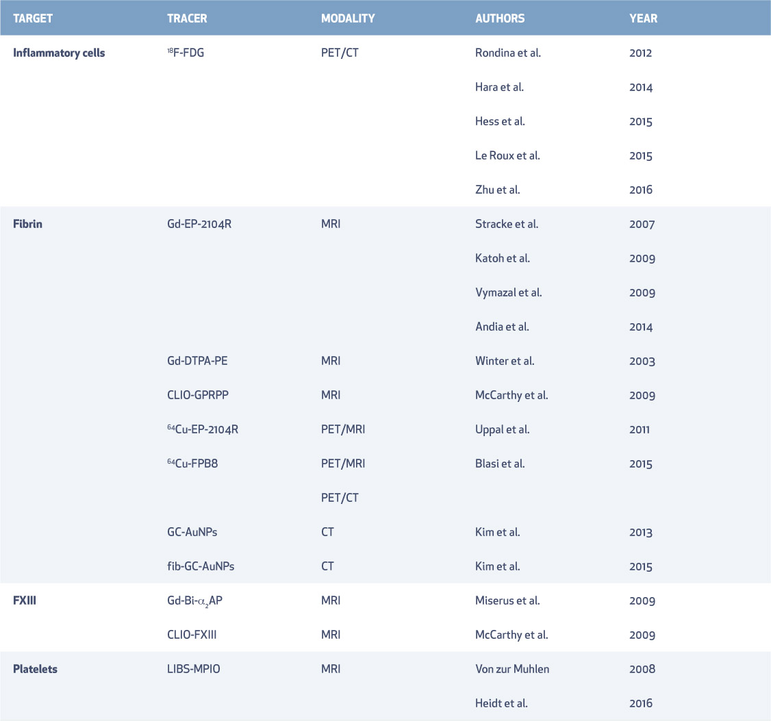
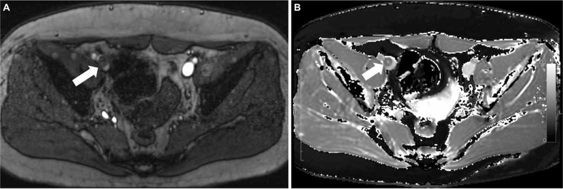
Figure 2.
(A) Time-of-flight magnetic resonance venography demonstrating right external iliac vein thrombus (white arrow) and (B) corresponding T1 map demonstrating low T1 relaxation time of the thrombus (white arrow).
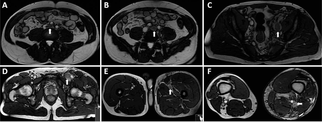
Figure 3.
Balanced steady-state free precession magnetic resonance venography of acute deep vein thrombosis. Thrombus is demonstrated by a white arrow in the inferior vena cava (A), common iliac vein (B), external iliac vein (C), common femoral vein (D), femoral vein (E), and popliteal vein (F).
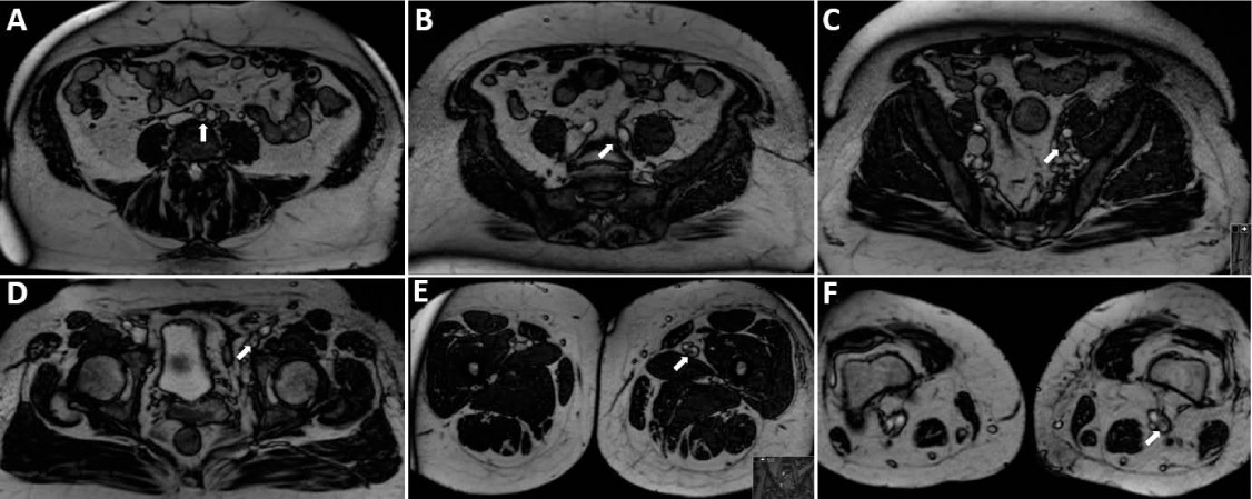
Figure 4.
Balanced steady-state free precession magnetic resonance venography of chronic deep vein thrombosis. Chronic changes (stenosed small-caliber vessels with post-thrombotic webs) are demonstrated by a white arrow in the inferior vena cava (A), common iliac vein (B), external iliac vein (C), common femoral vein (D), femoral vein (E), and popliteal vein (F).