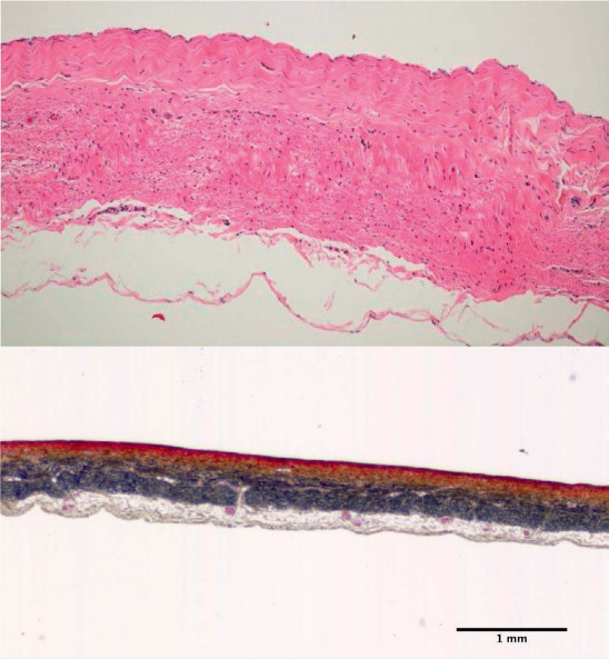
Figure 1.
The layered composition of the venous wall (intima is oriented to face top) is shown using hematoxylin and eosin (H&E) (top) and Movat pentachrome staining (bottom). The H&E staining shows the circumferentially aligned layers of protein fibers in the media distinct from the longitudinally aligned protein fibers in the adventitia. In the Movat-stained image, smooth muscle cells are stained red, collagen is yellow, elastin is black, and proteoglycans/glycosaminoglycans (PGs/GAGs) are blue. The media has dense smooth muscle cells arranged circumferentially between sheets of collagen and elastin. The thicker adventitia has dense sheets of longitudinally-aligned elastic fibers and collagen. PGs/GAGs are dispersed throughout the layers as well.