
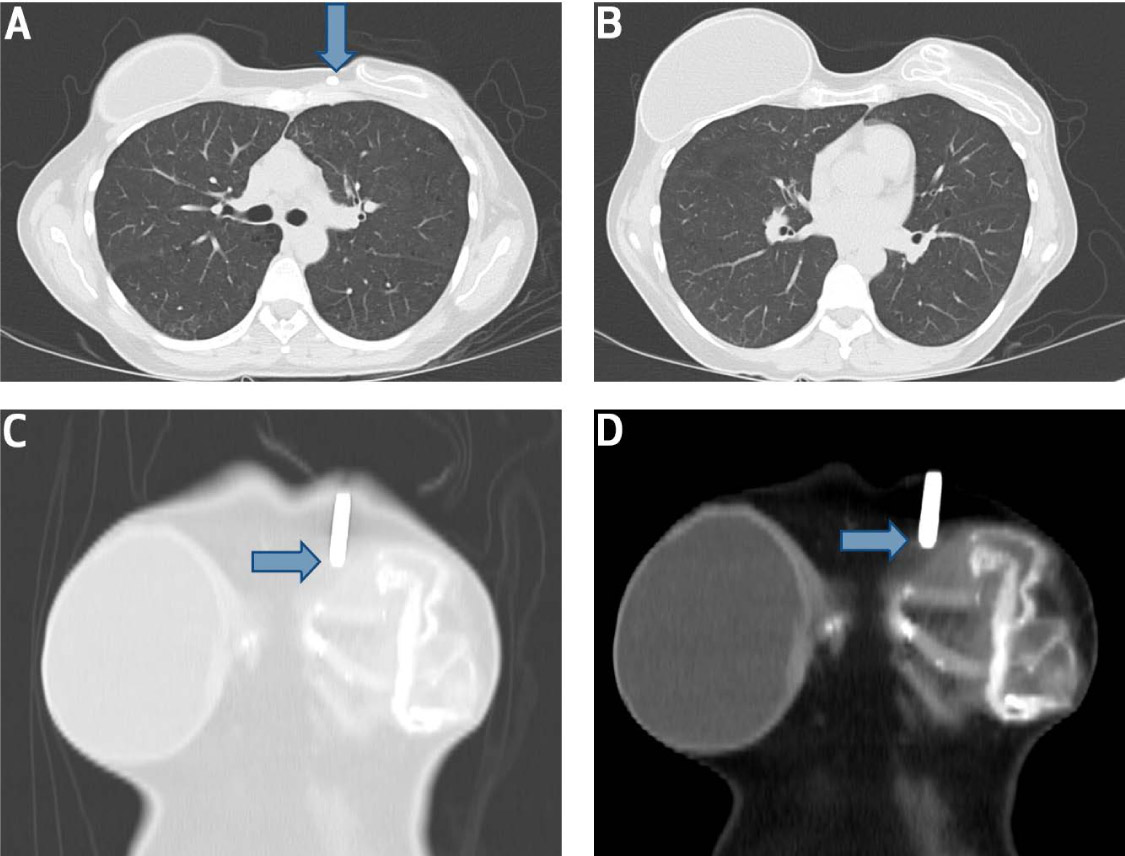
Figure 1.
Computed tomography scan of the chest (axial view) in a patient with subacute left breast implant rupture. (A) An insertable loop recorder (ILR) adjoins the site of left breast implant rupture (blue arrow). (B) Axial view of chest shows a clear difference in the morphology of the two breast implants. (C, D) Frontal views reveal close contact of the IRL (blue arrows) with the ruptured left breast implant contour.