
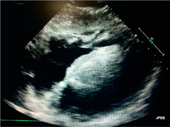
Figure 1.
Transthoracic echocardiogram showing severely enlarged interventricular septum with a thickness of 3.5 cm (normal range 0.7–1.1 cm).
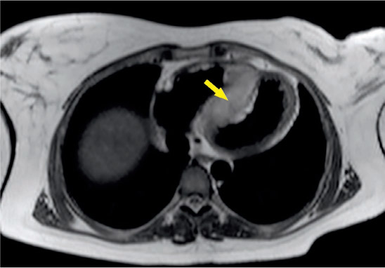
Figure 2.
Cardiac magnetic resonance imaging demonstrates a mass centered in the interventricular septum (arrow).
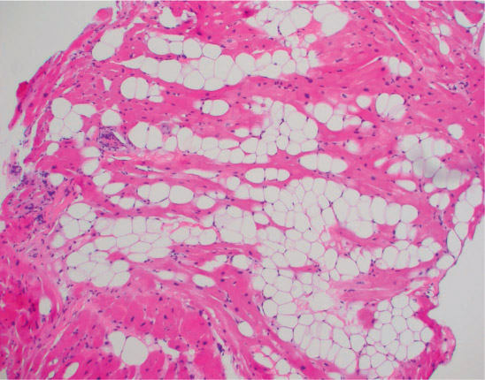
Figure 3.
Endomyocardial biopsy (H&E stained sections at 100× magnification), which illustrates abundant benign adipocytes transecting myocytes. The myocytes themselves appear relatively normal without hypertrophy or significant histologic findings.