
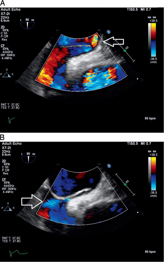
Figure 1.
Transesophageal echocardiography (TEE) still images. (A) Right-to-left flow across the patent foramen ovale (PFO) is demonstrated in early ventricular systole (arrow). (B) Left-to-right flow across the PFO is demonstrated in late ventricular systole (arrow).
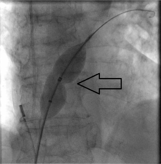
Figure 2.
Amplatzer sizing balloon in place across the patent foramen ovale. The waist (arrow) measured 13 mm.
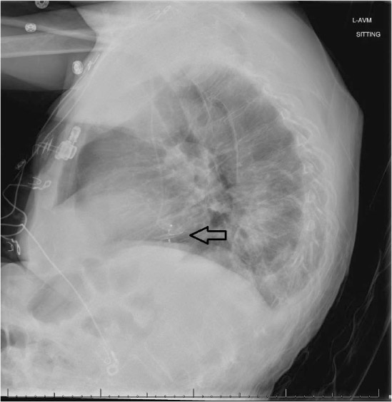
Figure 3.
Significant kyphosis on postprocedural lateral chest film. Note the presence of the Amplatzer device (arrow).