
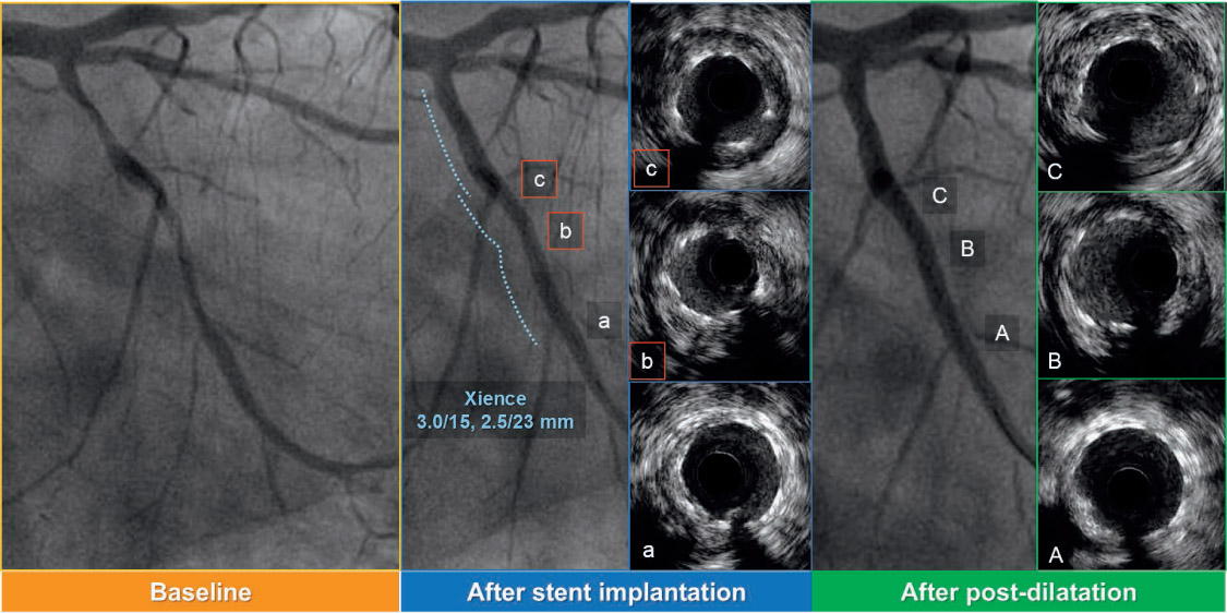
Figure 1.
Intravascular ultrasound (IVUS)-guided post-dilatation for the stent underexpansion. (a–c) IVUS images after stent implantation (b and c show stent underexpansion). Stent area (SA) is 3.45 mm2 in panel b, 3.81 mm2 in panel c. (A–C) IVUS images after IVUS-guided post-dilatation with 2.5-mm noncompliant balloon. SA: 4.65 mm2 in panel B, 6.03 mm2 in panel C.
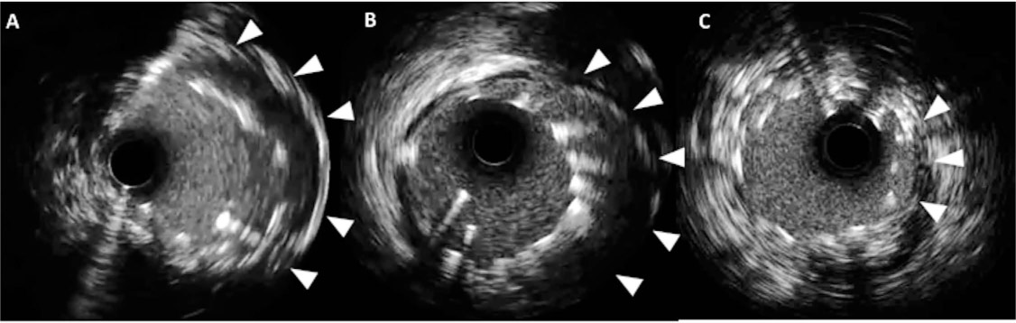
Figure 2.
Three cases of stent malapposition with intravascular ultrasound. (A–C) Substantial malapposition (arrowheads) is identified between vessel wall and stent struts.
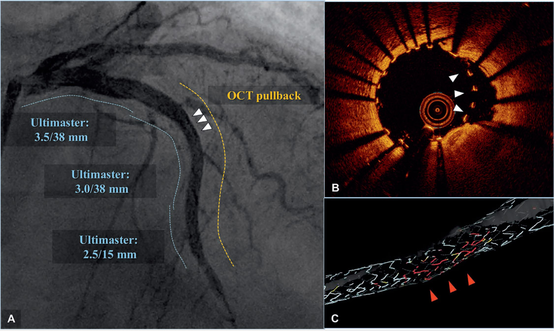
Figure 3 and Online Video 1.
Automatic stent struts detection system (OPTISTM Stent Optimization Software) of optical coherence tomography (OCT). Angiographic results (A) were excellent after implantation of an Ultimaster (Terumo Corp.) drug-eluting stent for a diffusely diseased left anterior descending artery (dotted lines indicate DES-treated area before [blue] and after OCT pullback [yellow]). However, despite the excellent angiographic results, OCT (B) showed malapposed struts (white arrowheads in 3 A and B), which were automatically detected and highlighted with red in a 3-dimensional reconstruction image (C).
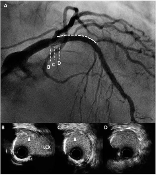
Figure 4.
(A) Coronary angiography (CAG) demonstrates dissection from the proximal edge of the stent (dotted line and D) to the left main trunk (indicated as B, C). (B–D) Cross-sectional intravascular ultrasound images show medial dissection (arrowheads) close to left circumflex artery (LCX).
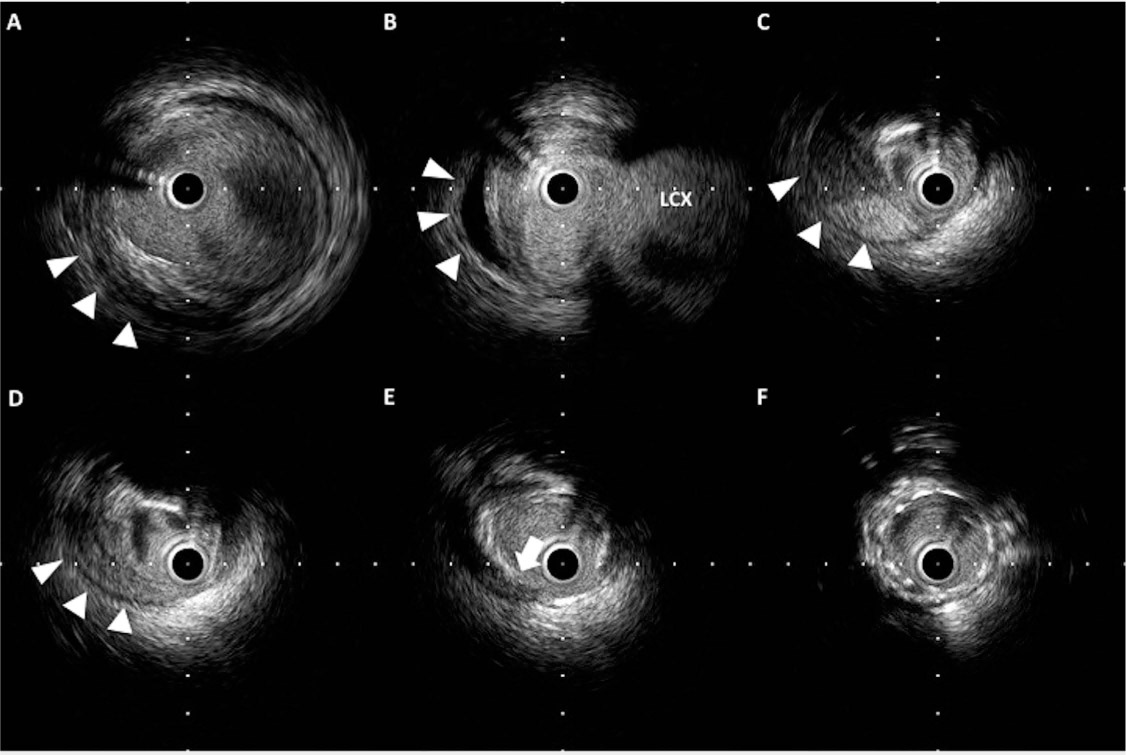
Figure 5.
Retrograde dissection and intramural hematoma. (A–D) Hematoma is observed within the media (arrowheads) and has pushed the lumen inward. (E) The entry site of the hematoma (arrow) reaches the media. (F) Stent is implanted with good apposition.
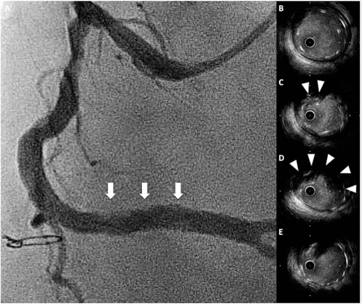
Figure 6.
Tissue protrusion. (A) Coronary angiography demonstrates a hazy appearance within the stent (arrows). (C, D) Cross-sectional IVUS images show tissue protrusion into the stent struts (arrowheads). (B, E) Stent is well-expanded without tissue protrusion.
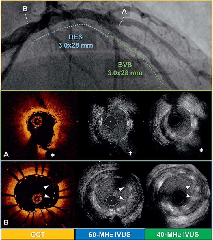
Figure 7.
Comparison of different intravascular imaging modalities (OCT, 40-MHz IVUS, and 60-MHz IVUS) after left main trunk (LMT) to left anterior descending artery (LAD) percutaneous coronary intervention with drug-eluting stent (DES) and bioresorbable vascular scaffold (BVS). (A) Mid-LAD lesion treated with BVS (asterisks indicate septal branch). (B) LMT lesion treated with DES (arrowheads indicate malapposed struts). Top panel shows final coronary angiography after the procedure (dashed lines indicate lesions treated with BVS [green] and DES [blue]).