
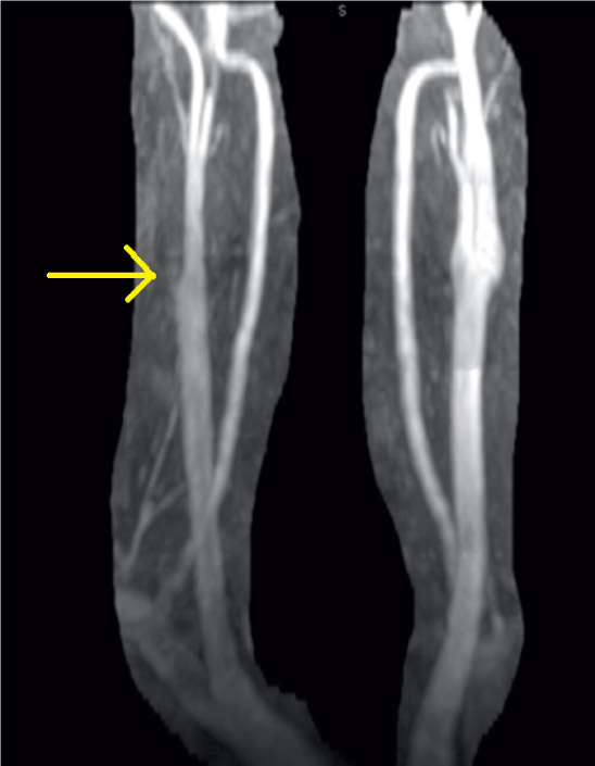
Figure 1.
Internal carotid artery occlusion on neck magnetic resonance angiography (arrow).
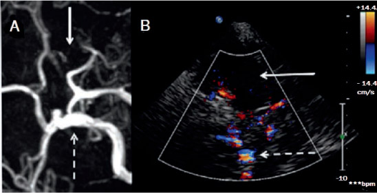
Figure 2.
Right internal carotid artery (ICA) occlusion (A) on magnetic resonance angiography and (B) on transcranial color-coded Doppler. Arrow: absent ICA
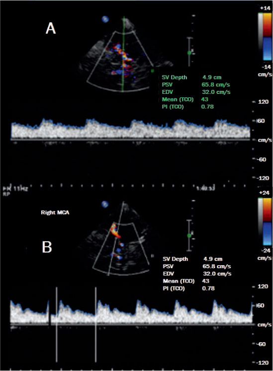
Figure 3.
(A) Blunted middle cerebral artery signal at 49-mm depth (baseline on 8.09.2012) and (B) normal waveform with straight systolic upstroke (post-recanalization on 9.04.2012).
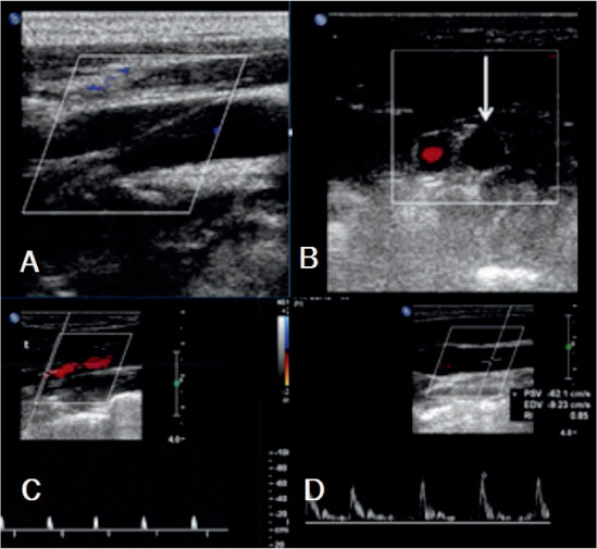
Figure 4.
(A) Right internal carotid artery (ICA) occlusion longitudinal view, (B) cross-sectional view (white arrow: absent color Doppler signal of the ICA), (C) ICA spectra waveform (high-resistance spikes to/from waveforms in the ICA), and (D) high-resistance waveform in the common carotid artery.
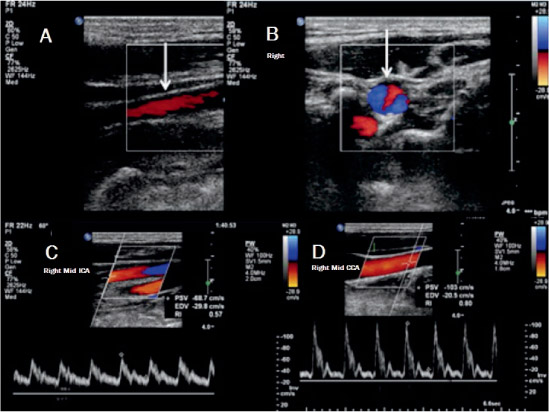
Figure 5.
(A) Internal carotid artery (ICA) recanalization longitudinal view (color Doppler signal present in ICA), (B) cross-sectional view (color Doppler signal present in ICA), (C) ICA spectra waveform (low-resistance waveform in the ICA), and (D) common carotid artery normal waveform.
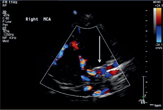
Figure 6.
Recanalized right internal carotid artery (arrow) on transcranial color-coded Doppler.
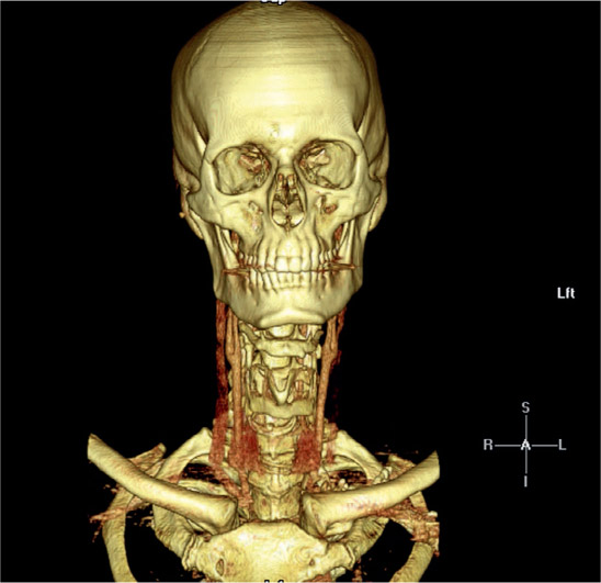
Figure 7.
Recanalization on computed tomography angiogram.