
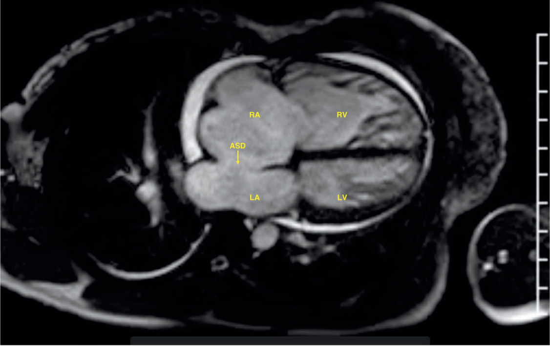
Figure 1.
Four-chamber view on cardiac magnetic resonance imaging showed a large 4-cm × 2.4-cm secundum atrial septal defect (ASD) with resultant severe right ventricular enlargement. RA: right atrium; LA: left atrium; RV: right ventricle; LV: left ventricle
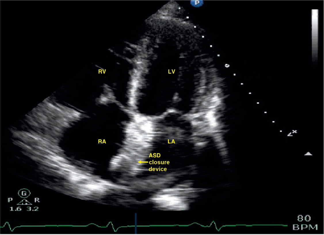
Figure 2.
Four-chamber view on a postprocedure transthoracic echocardiogram showed the atrial septal defect (ASD) closure device in the appropriate position. RV: right ventricle; RA: right atrium; LV: left ventricle; LA: left atrium
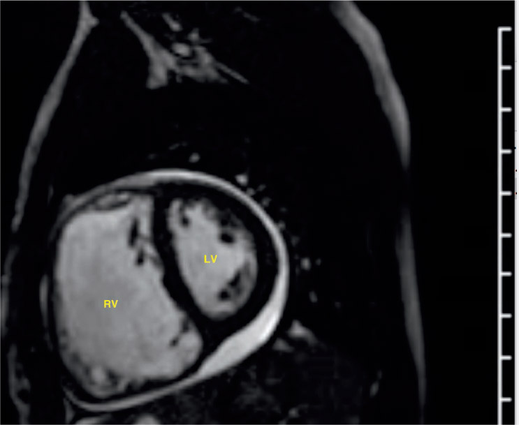
Figure 3.
D-shaped left ventricle (LV) and flattening of the interventricular septum on cardiac magnetic resonance imaging was consistent with right ventricular (RV) overload; significant RV overload likely led to LV failure due to ventricular interdependence.