Figure 1.
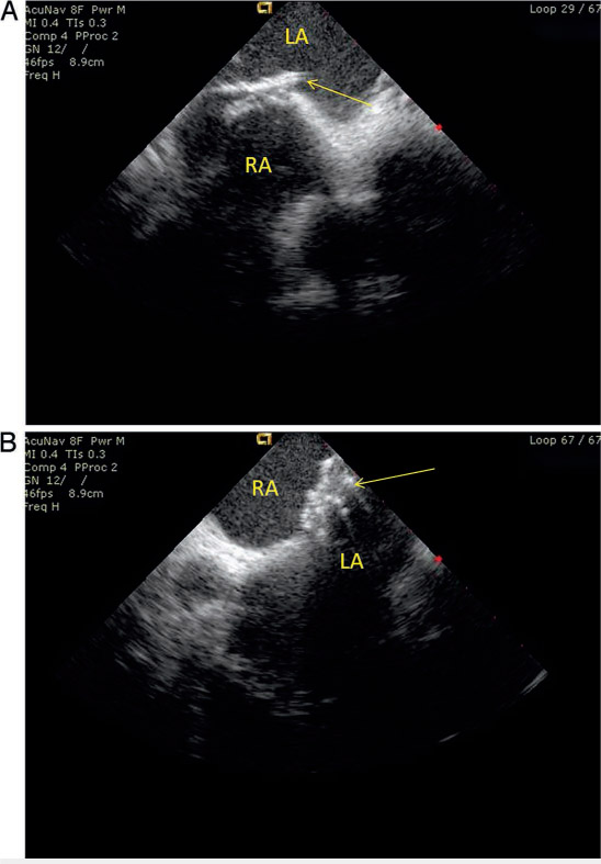
Closure device delivery. Intracardiac echocardiography showing (A) a delivery catheter placed across the PFO and (B) the Gore Helex Septal Occluder (W. L. Gore & Associates, Inc.) in appropriate position within the interatrial septum, sealing the PFO. RA: right atrium; LA: left atrium
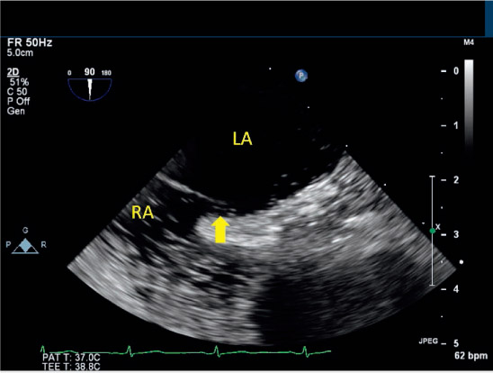
Figure 2.
Transesophageal echocardiogram (TEE) bubble study. Agitated saline bubbles directly visualized crossing the interatrial septum from right to left via a patent foramen ovale (arrow) using TEE in the bicaval view. LA: left atrium; RA: right atrium
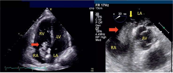
Figure 3.
Patent foramen ovale (PFO) with thrombus in transit. Intraoperative transesophageal echocardiogram showing a thrombus (red arrow) crossing the interatrial septum from right atrium (RA) to left atrium (LA) through a patent foramen ovale (yellow arrow). RV: right ventricle; LV: left ventricle; AV: aortic valve
Table 1.
Risk of Paradoxical Embolism calculator (RoPE). The above table shows the RoPE risk calculator for probability of paradoxical embolism as the etiology of cerebral infarction. The score ranges from 0 to 10, with higher scores indicating higher likelihood.13
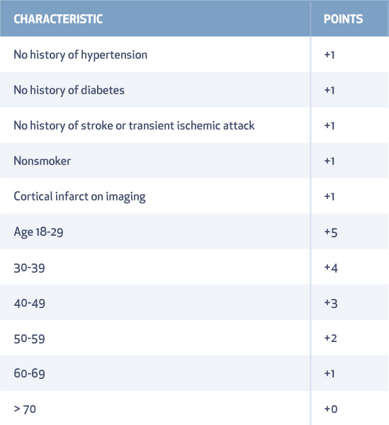
Table 2.
Patent foramen ovale (PFO) attributable risk percentage.13 RoPE: Risk of Paradoxical Embolism
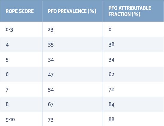
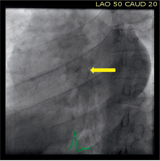
Figure 4.
This fluoroscopic image demonstrates the GORE CARDIOFORM Septal Occluder (yellow arrow) (W. L. Gore & Associates, Inc.) after deployment in the atrial septum.
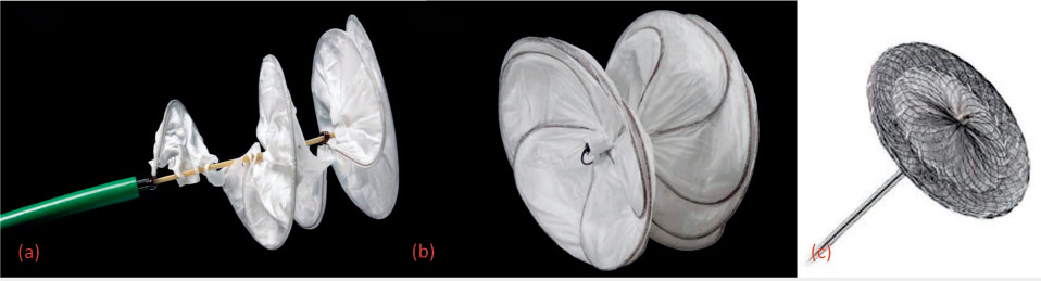
Figure 5.
Patent foramen ovale (PFO) occluder devices. This image shows (a) the GORE HELEX Septal Occluder (W. L. Gore & Associates, Inc.), (b) the GORE Cardioform Septal Occluder, and (c) the AMPLATZER PFO Occluder (St. Jude Medical).