
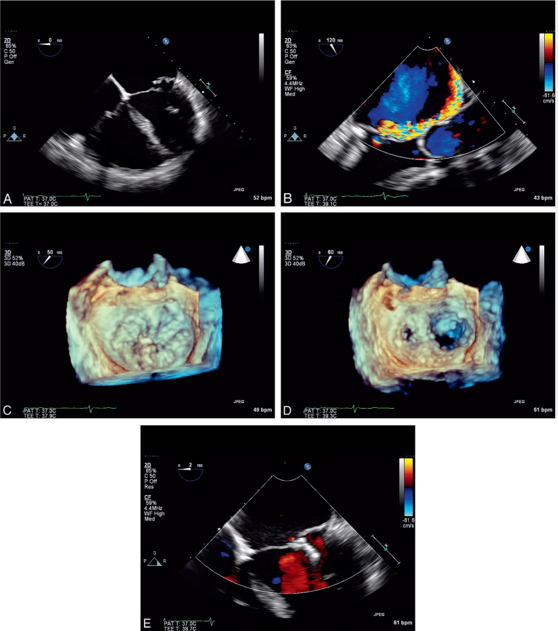
Figure 1.
Echocardiographic images of posterior mitral valve prolapse treated with percutaneous mitral valve repair using a MitraClip. (A) 2-dimensional (2D) echocardiogram showing posterior leaflet flail. (B) 2D echocardiogram with color Doppler showing significant mitral regurgitation. (C) 3D echocardiogram showing location of posterior leaflet flail. (D) 3D echocardiogram after grasp with mitral clip illustrating resultant double orifice mitral valve. (E) 2D echocardiogram with color Doppler showing significant reduction of mitral regurgitation after treatment with MitraClip.
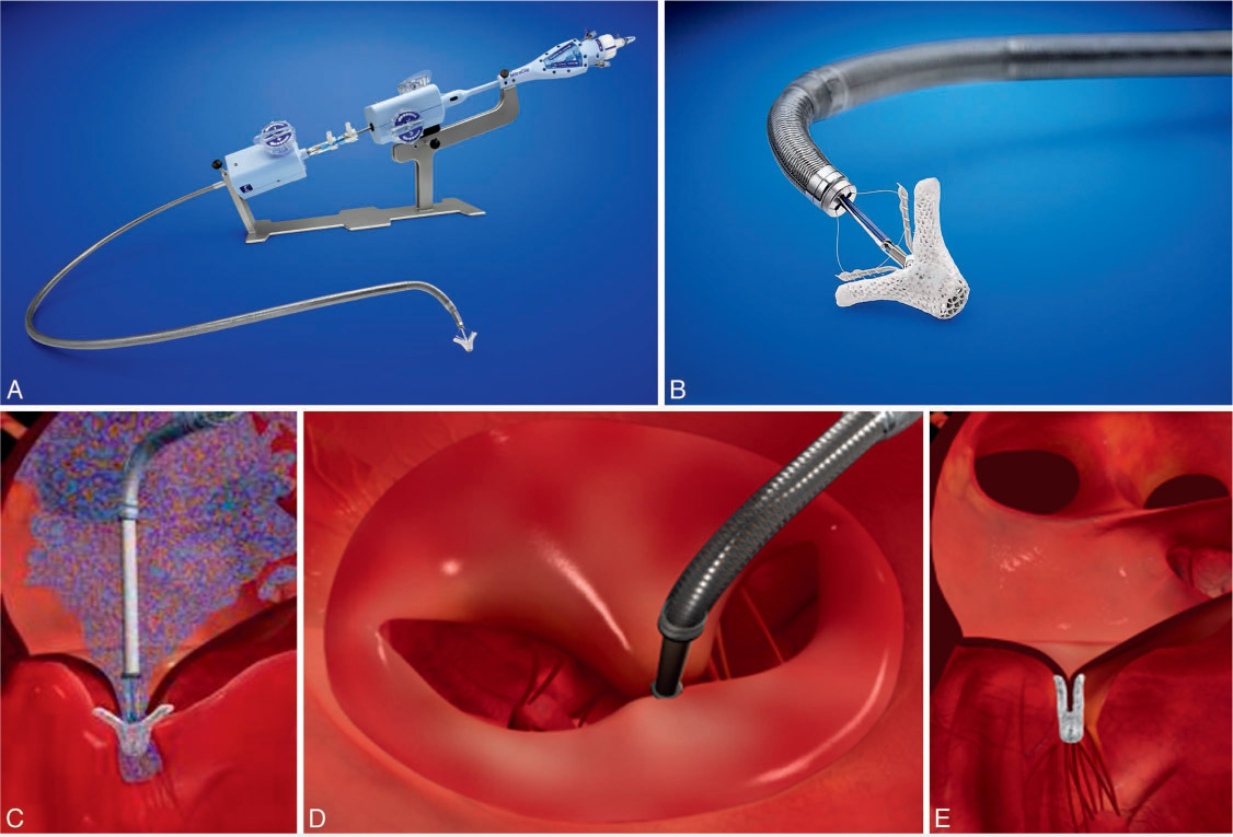
Figure 2.
Photographs and illustrations of the MitraClip System and procedure. (A) The MitraClip delivery system. (B) The MitraClip loaded on the delivery system open to 120 degrees with the grippers down. (C) Illustration of the MitraClip system engaging the mitral valve anterior and posterior leaflets at the site of the mitral regurgitation. (D) Illustration of the MitraClip grasping the mitral leaflets prior to being released. (E) Illustration of MitraClip after release.