
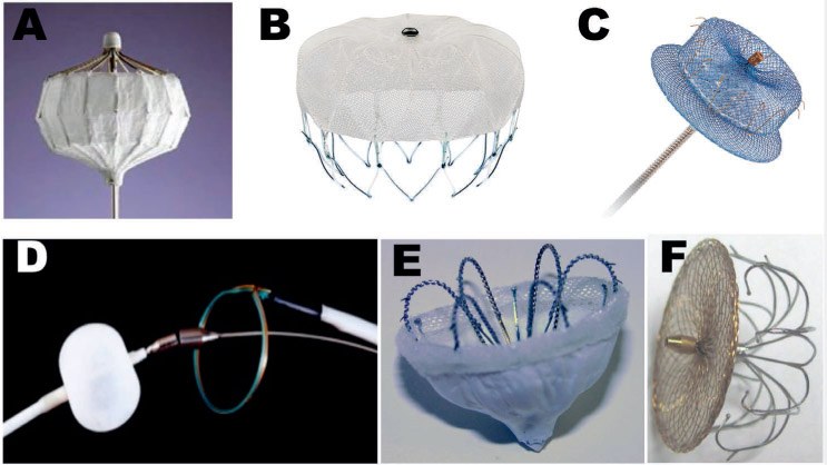
Figure 1.
Percutaneous devices for left atrial appendage occlusion. (A) PLAATO device; (B) WATCHMAN device; (C) Amplatzer Amulet device; (D) LARIAT suture delivery device; (E) WaveCrest device; (F) LAmbre device.
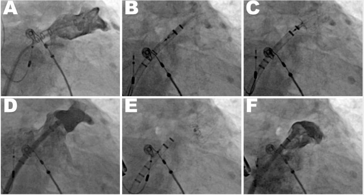
Figure 2.
Deployment of a WATCHMAN device. (A) Initial left atrial appendage (LAA) angiogram obtained through a pigtail catheter inserted via a sheath in the LAA. (B) Sheath advancement into the LAA with the WATCHMAN inside. (C) Deployment of the WATCHMAN in the LAA. (D) LAA angiogram to verify position of initial deployment in the LAA neck. (E) Release of the WATCHMAN. (F) Final angiogram.
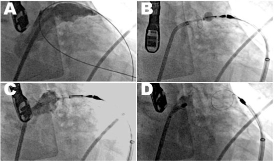
Figure 3.
Deployment of a LARIAT left atrial appendage (LAA) suture. (A) Initial LAA angiogram. (B) Endocardial and epicardial magnet-tipped wires snapped together. Balloon over endocardial wire aids in echocardiographic guidance. Snare has been advanced over the wires and is closed over the LAA. (C) Contrast injection in the left atrium shows complete occlusion. (D) After suture delivery, the snare is opened and retracted. Contrast injection shows a closed stump.