
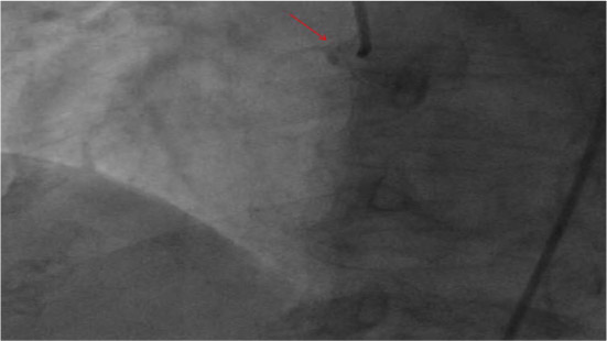
Figure 1.
Coronary angiogram showing a proximal right coronary artery (RCA) occlusion. See online video 1.
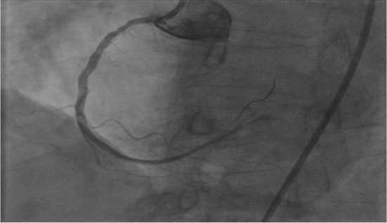
Figure 2.
Percutanous intervention with stent deployment in the proximal right coronary artery. See online video 2.
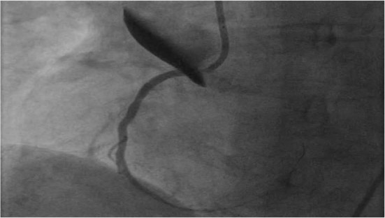
Figure 3.
Contrast injection after stent deployment resulting in acute dissection of the right coronary cusp with extension into the proximal ascending aorta. See online video 3.
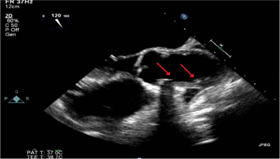
Figure 4.
Transesophageal echocardiogram showing an ascending aortic intramural hematoma (red arrows).
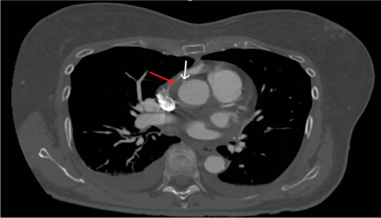
Figure 5.
Computed tomography (CT) scan showed a small aortic intimal tear (white arrow) with an associated intramural hematoma (red arrow).