

Have a personal or library account? Click to login
Pet Imaging and its Application in Cardiovascular Diseases Cover

Pet Imaging and its Application in Cardiovascular Diseases

Open Access
|Jan 2017

Figures & Tables

Table 1.

Imaging modality characteristics.8

i1947-6094-13-1-29-t01.jpg
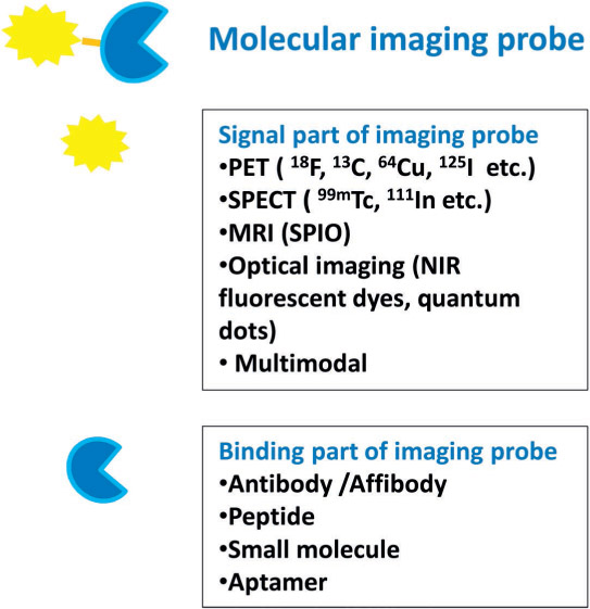
Figure 1.

Illustration of molecular imaging probe. PET: positron emission tomography; SPECT: single-photon emission computed tomography; MRI: magnetic resonance imaging; SPIO: superparamagnetic iron oxide; NIR: near infrared
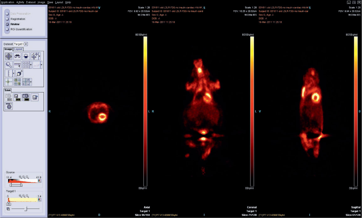
Figure 2.

Positron emission tomography images showing FDG uptake in the mouse heart. FDG: [18F]-2-fluoro-2-deoxy-D-glucose

Language: English
Page range: 29 - 33
Published on: Jan 1, 2017
Published by: Houston Methodist DeBakey Heart & Vascular Center
In partnership with: Paradigm Publishing Services

© 2017 Zheng Li, Anisha A. Gupte, Anjun Zhang, Dale J. Hamilton, published by Houston Methodist DeBakey Heart & Vascular Center
This work is licensed under the Creative Commons Attribution-NonCommercial 4.0 License.